Scanning mechanisms for imaging probe

Active Publication Date: 2008-07-24
SUNNYBROOK HEALTH SCI CENT
View PDF57 Cites 186 Cited by
  • Summary
  • Abstract
  • Description
  • Claims
  • Application Information

AI Technical Summary

Benefits of technology

[0008]A catheter-based system for intravascular ultrasound is described by Yock (U.S. Pat. No. 4,794,931) to provide high resolution imaging of structures in blood vessels. This system comprises an outer sheath, within which there is an ultrasound transducer near the distal end of a long torque cable. When a motor rotates the torque cable and ultr

Problems solved by technology

Higher frequencies provide higher resolution but result in lesser signal penetration and thus a smaller field of view.
However, optical coherence tomography is limited by its small penetration depth

Method used


Image

  • Scanning mechanisms for imaging probe
  • Scanning mechanisms for imaging probe
  • Scanning mechanisms for imaging probe

Examples


Example

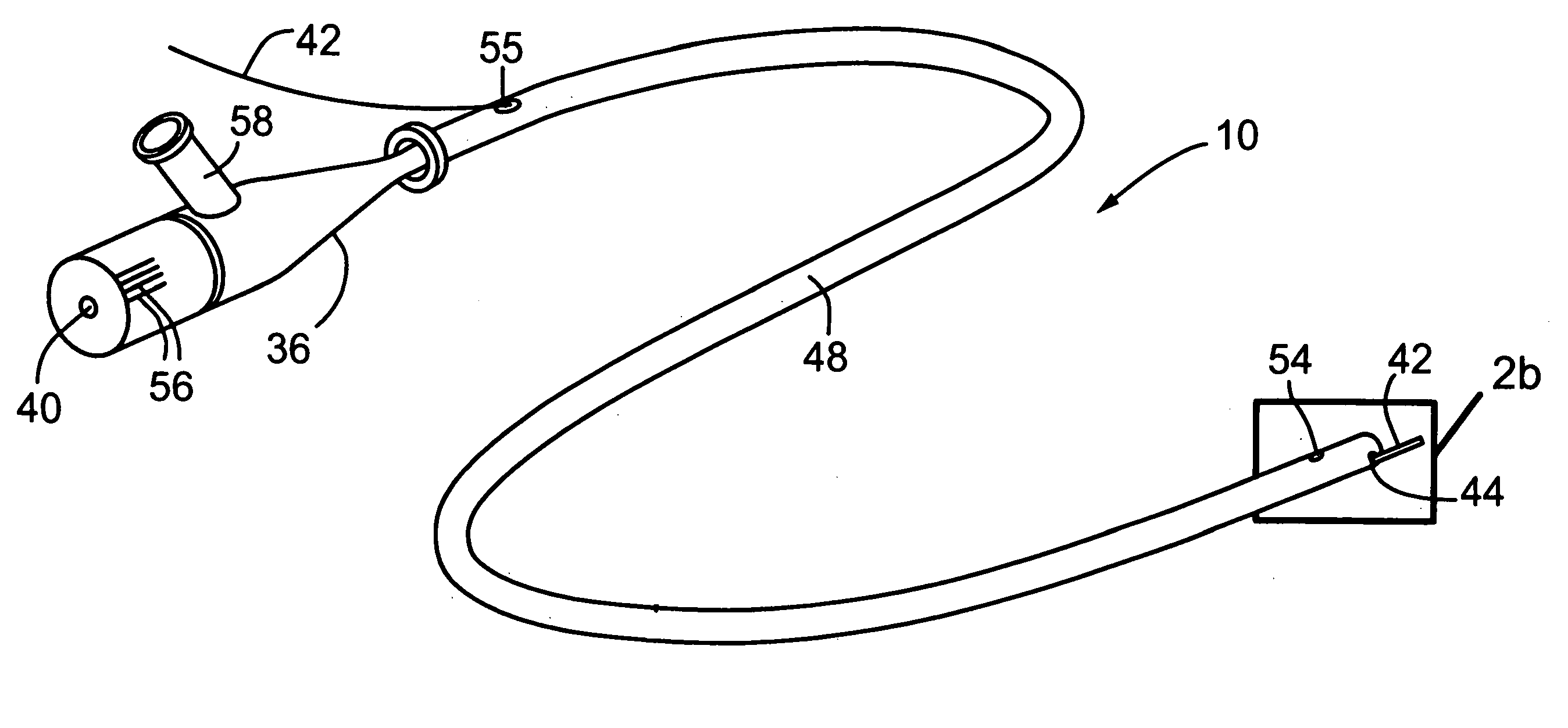
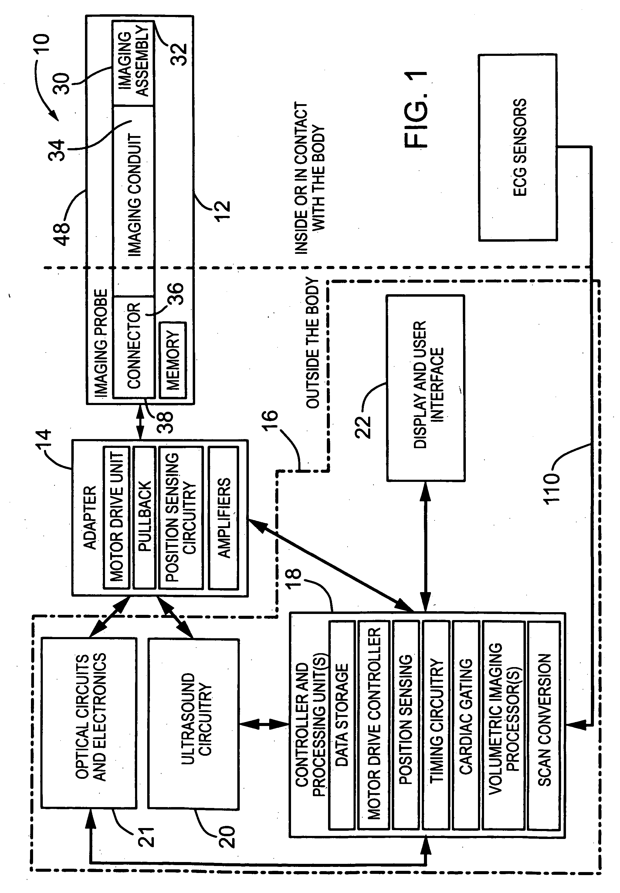
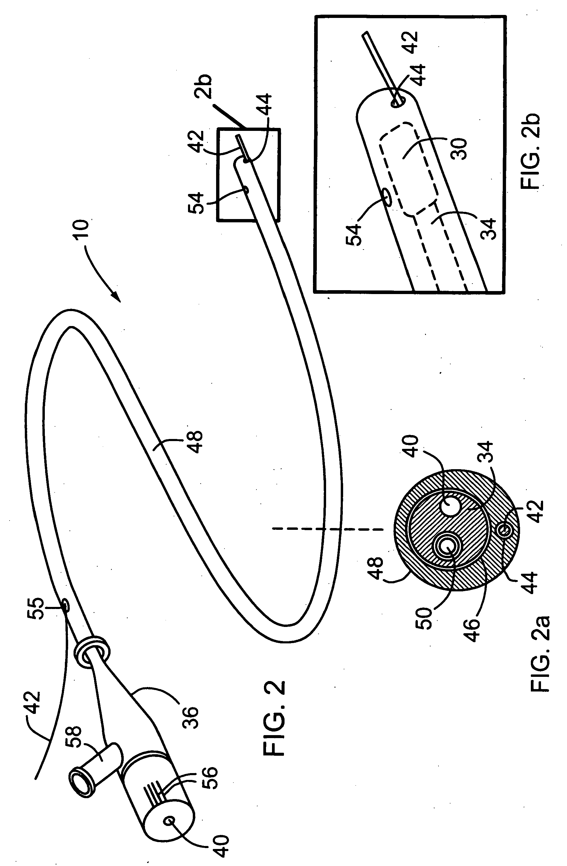
[0080]Generally speaking, the systems described herein are directed to an imaging probe using either optical or ultrasonic (or both) imaging. As required, embodiments of the present invention are disclosed herein. However, the disclosed embodiments are merely exemplary, and it should be understood that the invention may be embodied in many various and alternative forms. The Figures are not to scale and some features may be exaggerated or minimized to show details of particular elements while related elements may have been eliminated to prevent obscuring novel aspects. Therefore, specific structural and functional details disclosed herein are not to be interpreted as limiting but merely as a basis for the claims and as a representative basis for teaching one skilled in the art to variously employ the present invention. For purposes of teaching and not limitation, the illustrated embodiments are directed to an imaging probe.

[0081]As used herein, the terms “about”, and “approximately” ...


PUM


Abstract

The present invention provides scanning mechanisms for imaging probes using for imaging mammalian tissues and structures using high resolution imaging, including high frequency ultrasound and/or optical coherence tomography. The imaging probes include adjustable rotational drive mechanism for imparting rotational motion to an imaging assembly containing either optical or ultrasound transducers which emit energy into the surrounding area. The imaging assembly includes a scanning mechanism having including a movable member configured to deliver the energy beam along a path out of said elongate hollow shaft at a variable angle with respect to said longitudinal axis to give forward and side viewing capability of the imaging assembly. The movable member is mounted in such a way that the variable angle is a function of the angular velocity of the imaging assembly.

Description

CROSS REFERENCE TO RELATED U.S. APPLICATIONS[0001]This patent application relates to, and claims the priority benefit from, U.S. Provisional Patent Application Ser. No. 60 / 881,169 filed on Jan. 19, 2007, in English, entitled IMAGING PROBE, which is incorporated herein by reference in its entirety.FIELD OF THE INVENTION[0002]The present invention relates generally to the field of imaging probes for imaging mammalian tissues and structures using high resolution imaging, including high frequency ultrasound and optical coherence tomography. More particularly the present invention relates to imaging assemblies incorporating scanning mechanisms for providing forward and side viewing capabilities of the imaging probe.BACKGROUND OF THE INVENTION[0003]High resolution imaging of the body serves multiple purposes, including any of i) assessing tissue structures and anatomy; ii) planning and / or guiding interventions on localized regions of the body; and iii) assessing the result of intervention...

Claims


Application Information

Patent Timeline
IPC IPC(8): A61B1/04
CPCA61B5/0062A61B5/742A61B5/0095A61B5/6852A61B8/12A61B8/4281A61B8/4461A61B8/5238A61B8/543A61B1/00172A61B1/00183A61B8/483A61B8/445A61B5/0035A61B8/5292A61B5/0084A61B5/7207A61B5/0066G10K11/002A61B2562/0233A61B1/00112G02B6/3604G02B23/2423A61B1/05A61B1/04
Inventor COURTNEY, BRIANMUNCE, NIGEL ROBERTTHIND, AMANDEEP SINGHYANG, VICTOR XIAO DONGFOSTER, FRANCIS STUART
Owner SUNNYBROOK HEALTH SCI CENT
Why Eureka
Try Eureka