Depositing radiation in heart muscle under ultrasound guidance

a technology of heart muscle and ultrasound guidance, applied in the field of heart treatment, can solve the problems of limiting the rate at which x-ray images are acquired, blood cannot efficiently empty out of the atria into the ventricles, radiation exposure of collateral tissues, etc., and achieve the effect of reducing arrhythmias and improving radiosurgical treatment of tissues

Inactive Publication Date: 2008-07-24
CYBERHEART
View PDF45 Cites 60 Cited by
  • Summary
  • Abstract
  • Description
  • Claims
  • Application Information

AI Technical Summary

Benefits of technology

[0009]The present invention generally provides improved medical devices, systems, and methods, particularly for treatment of moving tissues. The invention allows improved radiosurgical treatment of tissues of the heart, often enhancing the capabilities of existing robotic radiosurgical systems for targeting tissues of the heart to mitigate arrhythmias such as atrial fibrillation or the like.

Problems solved by technology

During atrial fibrillation, the blood is not able to empty efficiently from the atria into the ventricles with each heart beat.
While well suited for treatment of lung tissues and the like, existing systems used to verify target registration may also limit radiation exposure of collateral tissues and / or avoid delays in the procedure by limiting the rate at which x-ray images are acquired during treatment.
As several radiation-sensitive structures are in and / or near the heart, and as the treatment time for a single heart patient may be as long as 30 minutes or more, increasing the imaging rate and / or delaying the radiation beams when the target tissue is not sufficiently aligned may be undesirable in many cases.

Method used


Image

  • Depositing radiation in heart muscle under ultrasound guidance
  • Depositing radiation in heart muscle under ultrasound guidance
  • Depositing radiation in heart muscle under ultrasound guidance

Examples


Embodiment Construction

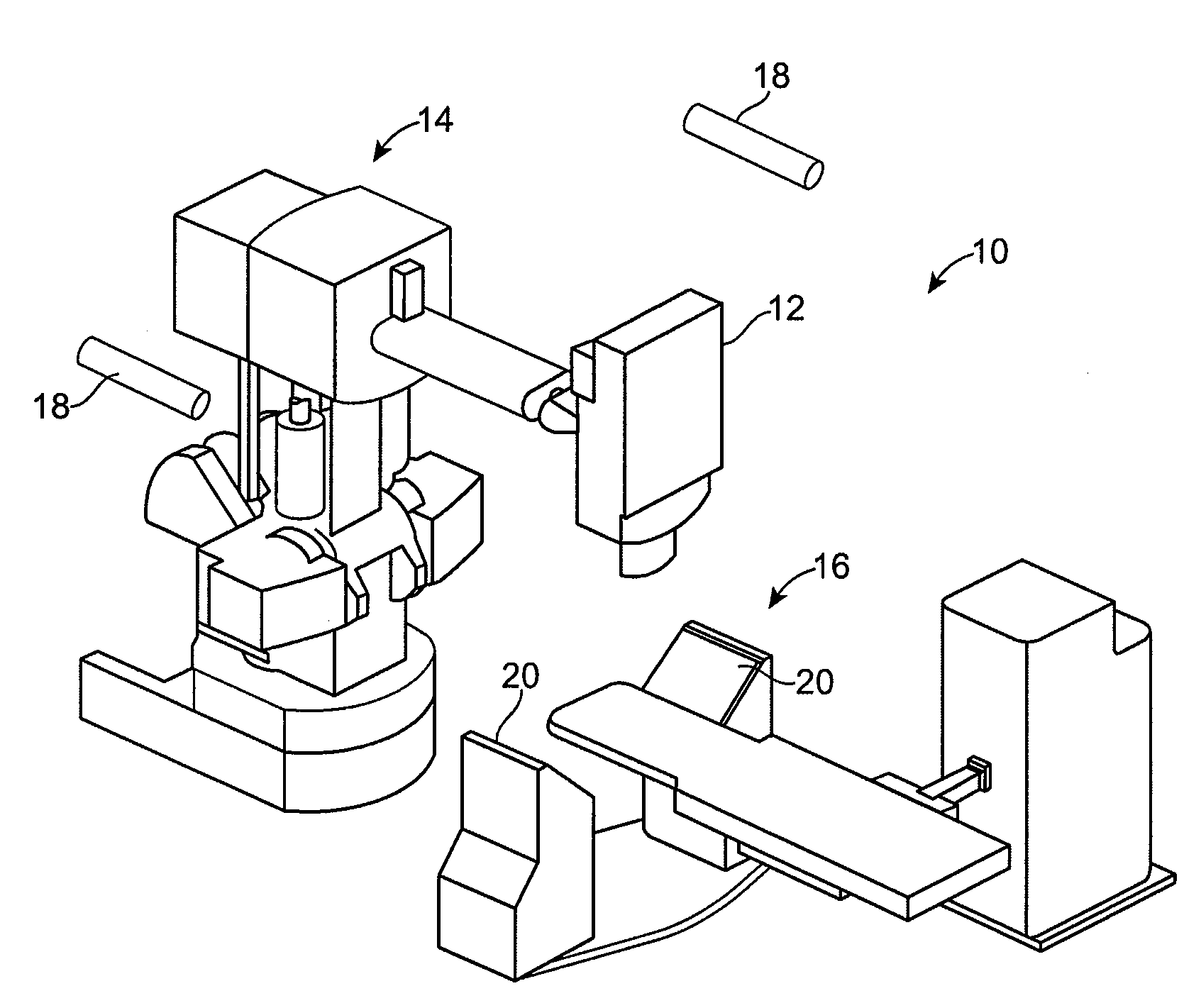
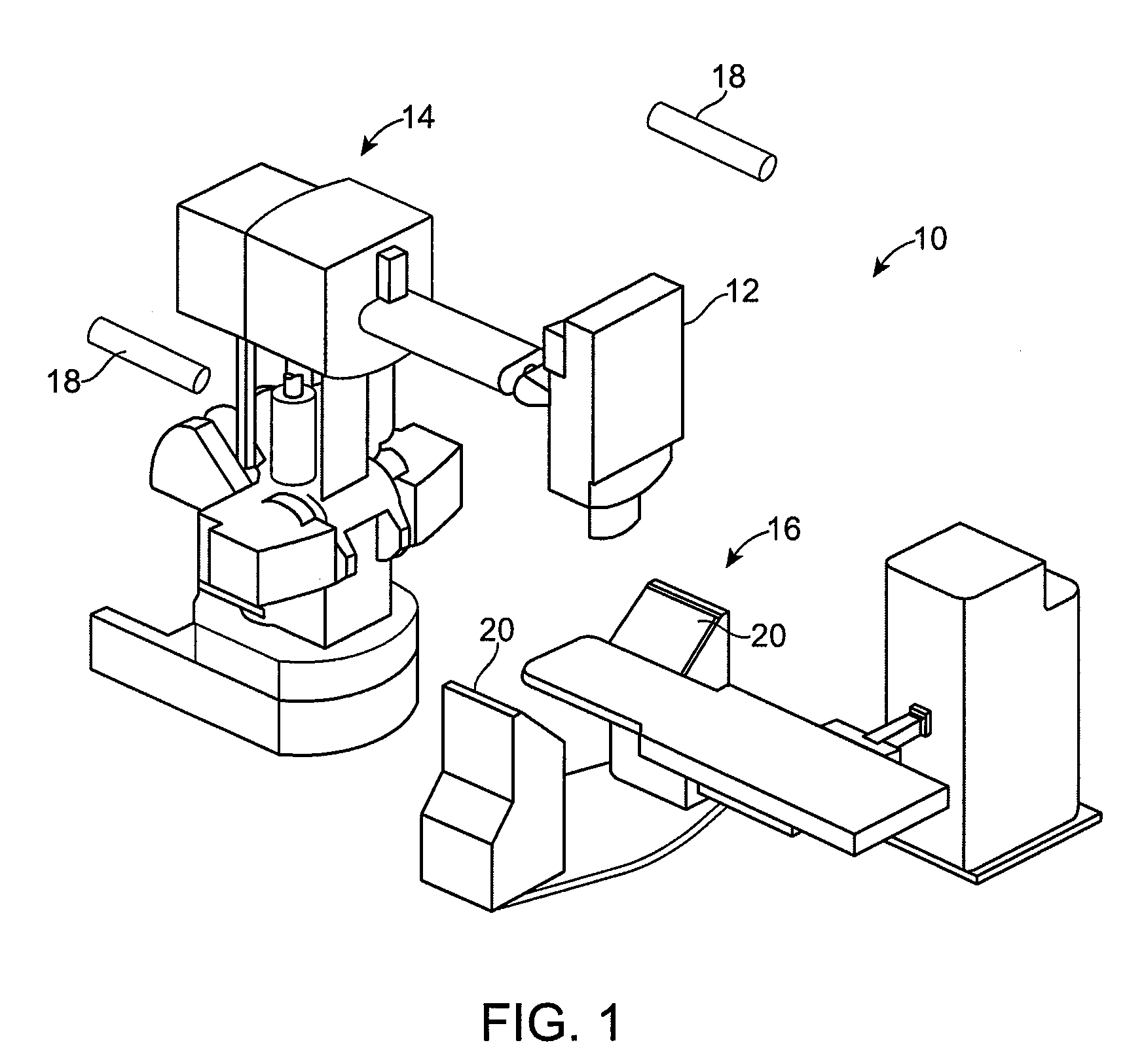
[0023]The present invention generally provides improved devices, systems, and methods for treatment of tissue, often using radiosurgical systems. The invention is particularly well suited for tracking of moving tissues such as tissues of the heart and tissue structures adjacent the heart that move with the cardiac or heartbeat cycles. The invention may take advantage of structures and methods which have been developed for treating tumors, particularly those which are associated with treatments of tissue structures that move with the respiration cycle. The cardiac cycle is typically considerably faster than the respiration cycle. The overall treatment times can also be quite lengthy for effective radiosurgical procedures on the heart (typically being greater than 10 minutes, often being greater than ½ hour, and in many cases, being two hours or more). Hence, it will often be advantageous to avoid continuous imaging of the target and adjacent tissues using fluoroscopy or the like. A v...


PUM


Abstract

A method and system are disclosed for radiosurgical treatment of moving tissues of the heart, including acquiring at least one volume of the tissue and acquiring at least one ultrasound data set, image or volume of the tissue using an ultrasound transducer disposed at a position. A similarity measure is computed between the ultrasound image or volume and the acquired volume or a simulated ultrasound data set, image or volume. A robot is configured in response to the similarity measure and the position of the transducer, and a radiation beam is fired from the configured robot.

Description

CROSS-REFERENCES TO RELATED APPLICATIONS[0001]This application claims the benefit under 35 USC 119(e) of U.S. Provisional Application No. 60 / 879,654, filed on Jan. 9, 2007, and entitled “Depositing Radiation In Heart Muscle Under Ultrasound Guidance”, and U.S. Provisional Application No. 60 / 879,724, filed on Jan. 9, 2007, entitled “Method For Depositing Radiation In Heart Muscle”, the full disclosures of which are incorporated herein by reference.[0002]This application is related to U.S. Provisional Application No. 60 / 918,540, filed on Mar. 16, 2007, entitled “Radiation Treatment Planning And Delivery For Moving Targets In The Heart”, and U.S. Provisional Application No. 60 / 975,373, filed on Sep. 26, 2007, entitled “Radiosurgical Ablation of the Myocardium”, the full disclosures of which are incorporated herein by reference.BACKGROUND OF THE INVENTION[0003]The present invention is generally related to treatment of the heart, and in particular, non-invasive treatment of the heart usi...

Claims


Application Information

Patent Timeline
Patent Type & Authority Applications(United States)
IPC IPC(8): A61B19/00
CPCA61B19/20A61N2005/1058A61B19/50A61B19/52A61B19/5244A61B2017/00044A61B2017/00243A61B2017/00703A61B2019/207A61B2019/2211A61B2019/505A61B2019/507A61B2019/5238A61B2019/524A61B2019/5255A61B2019/5276A61B2019/5287A61B2019/5289A61B2019/5445A61N5/1049A61N5/1067A61B19/2203A61B90/10A61B90/36A61B2034/2055A61B2090/363A61B2090/3945A61B34/20A61B2034/301A61B2090/364A61B2090/376A61B34/30A61B2090/101A61B34/10A61B2034/105A61B2034/107A61B2090/3762A61B2090/378A61N5/1037A61N5/1068
Inventor SUMANAWEERA, THILAKAMAGUIRE, PATRICK
Owner CYBERHEART
Why Eureka
Try Eureka