Methods of prenatal screening for trisomy 21

a trisomy 21 and prenatal screening technology, applied in the field of trisomy 21 prenatal screening, can solve the problems that the difference between maxillary measurements between trisomy 21 and euploid fetuses has not been large enough to be clinically useful, and the effect of reducing the risk of euploid fetuses and reducing the risk of euploid fetus developmen

Inactive Publication Date: 2008-08-07
SONEK JIRI D +1
View PDF0 Cites 25 Cited by
  • Summary
  • Abstract
  • Description
  • Claims
  • Application Information

AI Technical Summary

Benefits of technology

[0007]In one embodiment, the present invention is directed to a method for prenatal screening for trisomy 21, comprising obtaining a two or three dimensional image of a fetal face, measuring the angle between a line extending along the upper aspect of the maxilla and a line extending from the anterior aspect of the maxilla to the external surface of the frontal bone at the point...

Problems solved by technology

However, from these studies, the reported differences between maxillary measurements betwe...

Method used


Image

  • Methods of prenatal screening for trisomy 21
  • Methods of prenatal screening for trisomy 21
  • Methods of prenatal screening for trisomy 21

Examples


example 1

[0026]This retrospective study utilized 3D volumes of the fetal face, which are acquired before fetal karyotyping by chorionic villus sampling (CVS) at 11-13+6 weeks of gestation. The fetuses are determined as being at risk of trisomy 21 based on the combination of maternal age and nuchal translucency measurement. Singleton pregnancies only are used. In each fetus, the crown-rump length and nuchal translucency thickness are measured in a standard fashion. A mid sagittal image of the fetal face is examined for the presence or absence of the nasal bone.

[0027]A database of stored images and diagnoses is reviewed and 100 consecutive cases of fetal trisomy 21 and 300 chromosomally normal fetuses in which a 3D volume of the fetal face had been obtained, are identified. The images are obtained with the fetus in the mid-sagittal plane with the transducer being parallel to the long axis of the nose. All 3D examinations are carried out transabdominally (RAB 4-8L probe, Voluson 730 Expert, GE ...

example 2

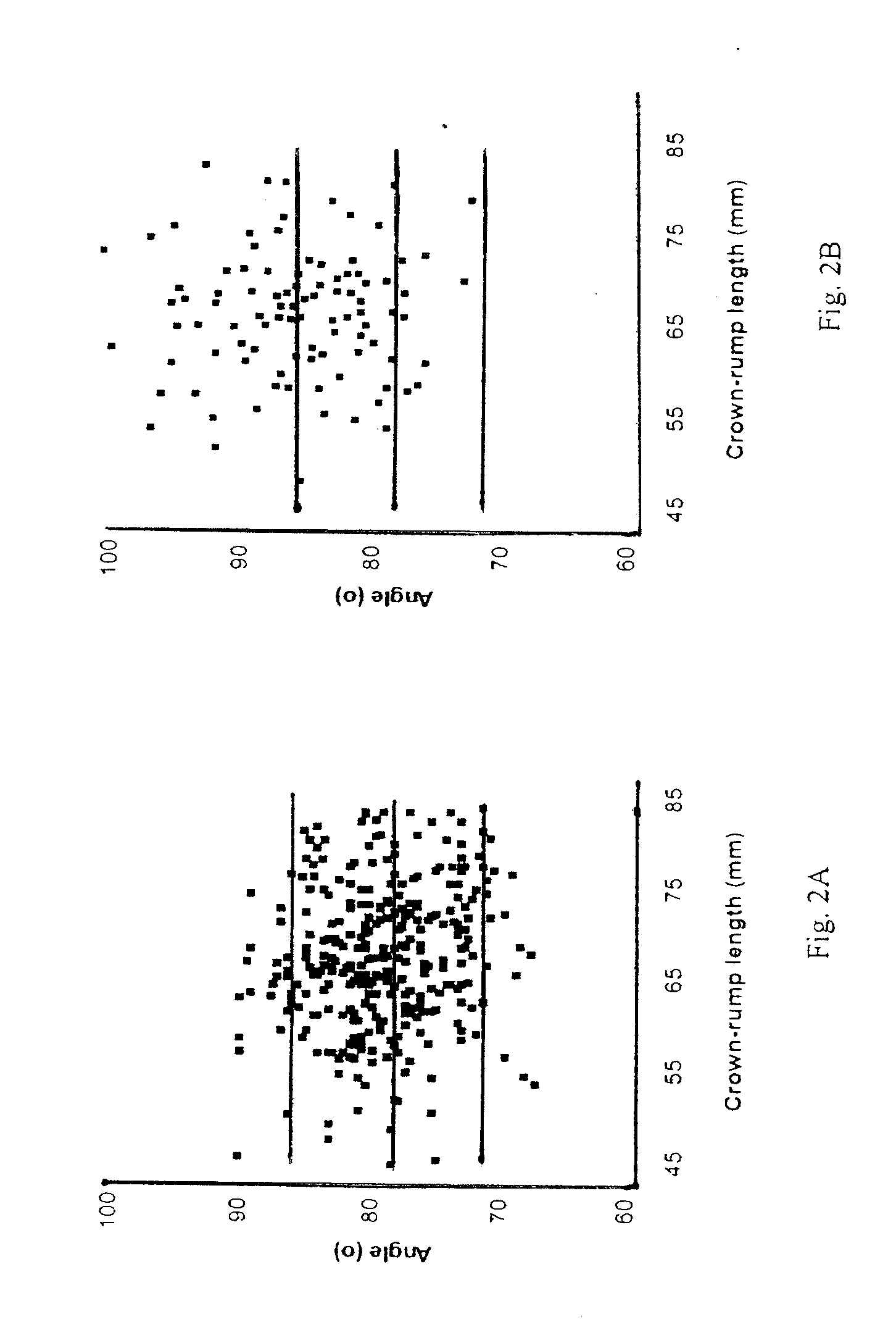
[0037]This study involved a review of stored digital 2D images of fetal profiles, taken from singleton pregnancies before amniocentesis for fetal karyotyping at 14-24 weeks of gestation, wherein 100 consecutive pregnancies in which the fetal karyotype was normal and 34 fetuses with trisomy 21 were examined. All images were obtained using Sequoia 512 (Siemens) system. The angles were measured using on-line tools in the KinetDx (Siemens) PAC system.

[0038]The following data were retrieved from the patient records: maternal age, gestational age, indication for karyotyping, the findings at the ultrasound examination carried out before amniocentesis, including fetal abnormalities or markers for aneuploidy and presence or absence of the nasal bone.

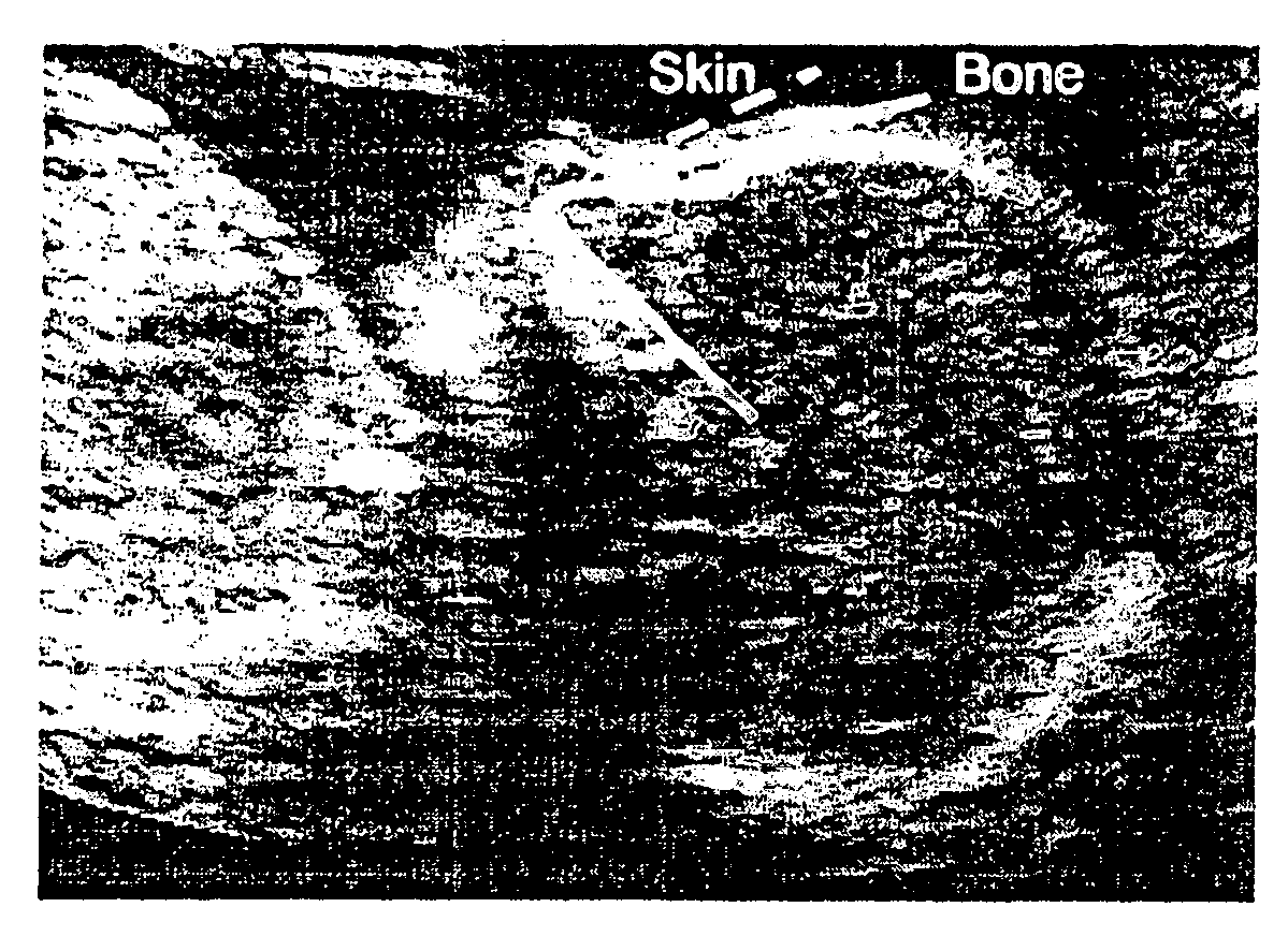
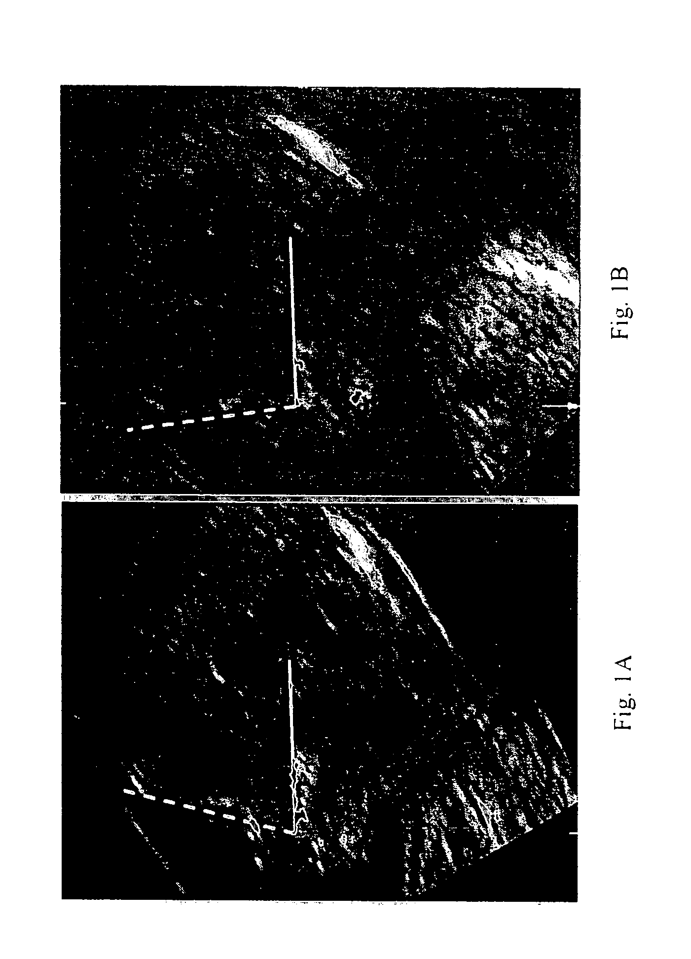
[0039]The images had been obtained by transabdominal sonography with the fetus facing the transducer. The ideal angle of insonation with respect to the longitudinal axis of the upper palate was determined to be about 45°. In this plane the upper ...


PUM


Abstract

Methods for prenatal screening for trisomy 21 employ examination of the fronto-maxillary facial (FMF) angle of a fetus. In one embodiment, the methods comprise obtaining a two or three dimensional image of a fetal face, measuring the FMFbone angle on the image, and comparing the measured FMFbone angle with an FMFbone angle characteristic of chromosomally normal fetuses, wherein a measured FMFbone angle greater than the FMFbone angle characteristic of chromosomally normal fetuses provides an indication of an increased likelihood of the occurrence of trisomy 21 in the fetus. In another embodiment, the methods comprise obtaining a two or three dimensional image of a fetal face, measuring the FMFskin angle on the image, and comparing the measured FMFskin angle with an FMFskin angle characteristic of chromosomally normal fetuses, wherein a measured FMFskin angle greater than the FMFskin angle characteristic of chromosomally normal fetuses provides an indication of an increased likelihood of the occurrence of trisomy 21 in the fetus. In additional embodiments, both FMFbone angle and FMFskin angle are measured and compared with values characteristic of chromosomally normal fetuses.

Description

CROSS-REFERENCES TO RELATED APPLICATIONS[0001]The present application claims the benefit under 35 U.S.C. § 119 of U.S. Application Ser. Nos. 60 / 834,579 filed Aug. 1, 2006 and 60 / 872,880 filed Dec. 5, 2006, both of which are incorporated herein by reference.FIELD OF THE INVENTION[0002]The present invention is directed to methods for prenatal screening for trisomy 21, the most common form of Down syndrome, by examination of the fronto-maxillary facial (FMF) angle of a fetus.BACKGROUND OF THE INVENTION[0003]Down syndrome is a genetic condition in which a person has 47 chromosomes instead of the usual 46. In most cases, Down syndrome occurs when there is an extra copy of chromosome 21. This form of Down syndrome is known as trisomy 21.[0004]Trisomy 21 is associated with a specific phenotype, which includes a flat profile. This observation is part of Langdon Down's original description of what later became known as Down syndrome. Several radiological studies have reported underdevelopmen...

Claims


Application Information

Patent Timeline
IPC IPC(8): A61B8/13A61B5/107
CPCA61B5/1075A61B8/483A61B8/0866A61B5/4504
Inventor SONEK, JIRI D.NICOLAIDES, KYPROS H.
Owner SONEK JIRI D
Why Eureka
Try Eureka