Methods for minimally invasive, non-permanent occlusion of a uterine artery

a uterine artery and non-permanent occlusion technology, applied in the field of non-permanent occlusion of the uterine artery or artery, can solve the problems of 0.5 death rate, difficult to know which fibroid is causing symptoms for the patient, and uterine fibroid disorders

Inactive Publication Date: 2008-08-21
VASCULAR CONTROL SYST
View PDF99 Cites 12 Cited by
  • Summary
  • Abstract
  • Description
  • Claims
  • Application Information

AI Technical Summary

Problems solved by technology

Hysterectomy for treating uterine fibroid disorders, though effective, has many undesirable characteristics.
The undesirable characteristics of hysterectomy include a known mortality rate of 0.5 deaths per 1000 hysterectomies.
Consequently, it is difficult to know which fibroid is causing symptoms to the patient (bleeding, pain, and bulk effects on adjacent organs).
Clearly, the strategy of identifying which individual fibroid is causing symptoms (when there are often many), finding that fibroid, and then either removing or destroying that individual fibroid is a rather complex strategy.
Second, fibroids live a tenuous vascular life with very little ability to recruit a new blood supply from the host when the primary blood supply is compromised.
This procedure has been performed laparoscopically and requires a great deal of surgical skill to access, identify, dissect, and ligate the uterine arteries.
This requirement for high skill and a full surgical approach has limited the use of surgical ligation of the uterine arteries as a clinical alternative for uterine fibroid treatment.
Heretofore, this inherent characteristic of these tissues has not been utilized in the treatment of myomata, or for accessing the uterine artery.
The difficulty and cost associated with the use of traditional transvascular access, and the possibility of infection and surgical complication associated with dissection, render these prior techniques unacceptable.
Post partum hemorrhaging and cesarean section-related bleeding can be a dangerous if not quickly and adequately controlled, which may require a fast surgical response, from which the patient may suffer from associated trauma of the surgery.
This ligation task can be very laborious and time-consuming, as the arteries are dissected and ligated by the surgeon, and have associated complications.

Method used


Image

  • Methods for minimally invasive, non-permanent occlusion of a uterine artery
  • Methods for minimally invasive, non-permanent occlusion of a uterine artery
  • Methods for minimally invasive, non-permanent occlusion of a uterine artery

Examples


Embodiment Construction

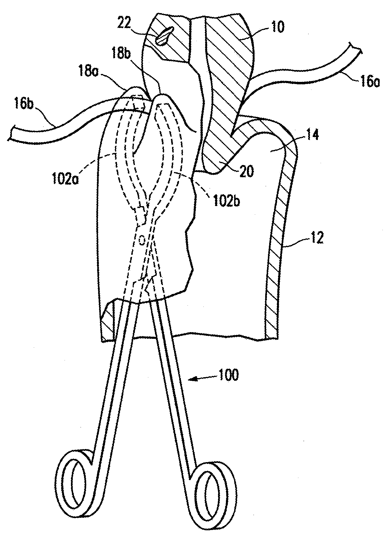
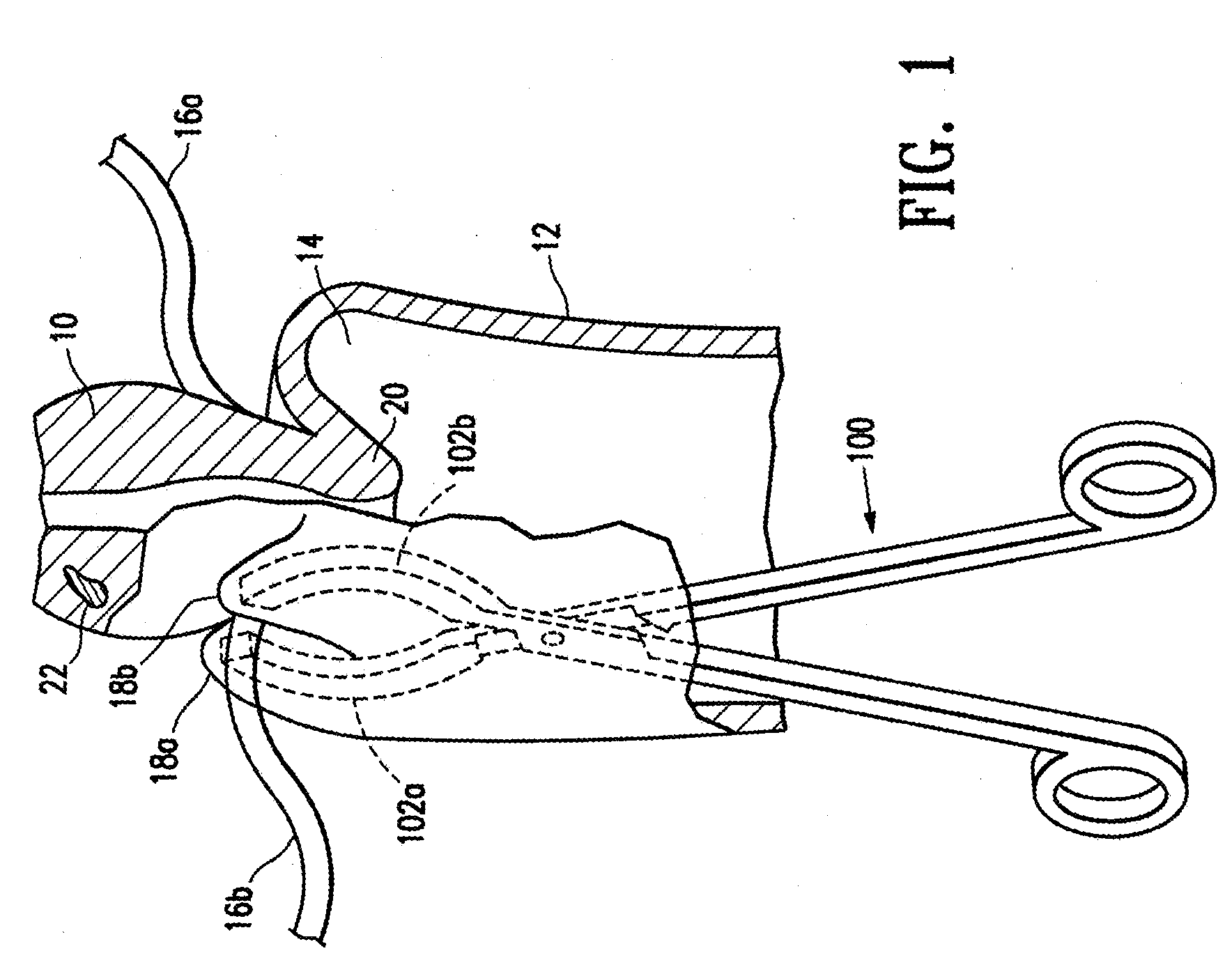
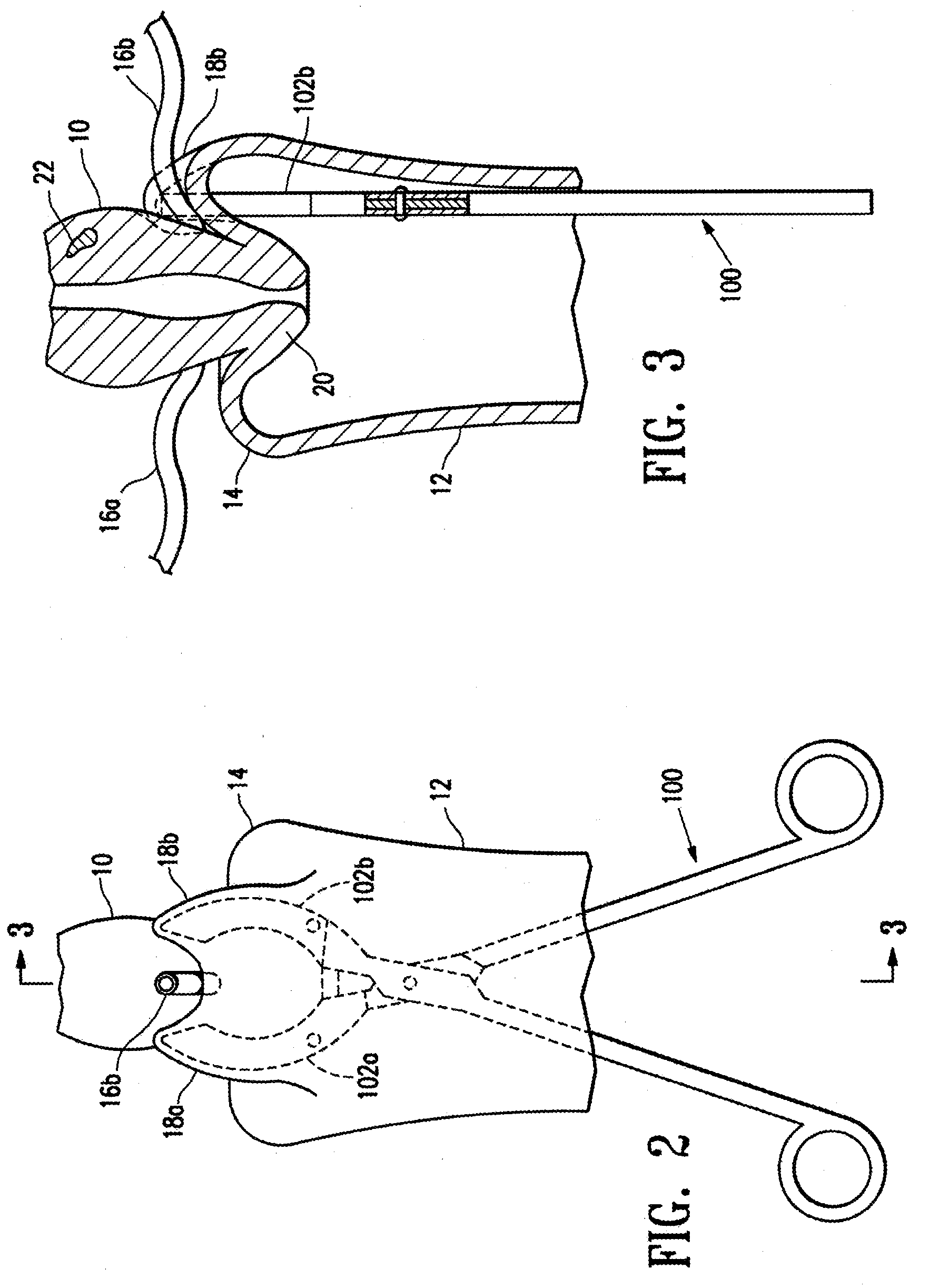
[0041]The headings used herein are for organizational purposes only and are not meant to be used to limit the scope of the description or the claims. As used throughout this application, the word “may” is used in a permissive sense (i.e., meaning having the potential to), rather than the mandatory sense (i.e., meaning must). Similarly, the words “include”, “including”, and “includes” mean including but not limited to. To facilitate understanding, like reference numerals have been used, where possible, to designate like elements common to the figures.

[0042]Non-permanent occlusion of the uterine artery is sufficient to cause the demise of uterine myomata without unnecessarily exposing other tissues and anatomical structures to hypoxia attendant to prior permanent occlusion techniques. Burbank, Fred, et al., Uterine Artery Occlusion by Embolization or Surgery for the Treatment of Fibroids: A Unifying Hypothesis-Transient Uterine Ischemia, The Journal of the American Association of Gyne...


PUM

PropertyMeasurementUnit
time periodaaaaaaaaaa
transient timeaaaaaaaaaa

Abstract

A method of treating a uterine disorder comprising non-permanently occluding a uterine artery with a resorbable embolic mass for a therapeutically effective time period. The occluding step includes selectively positioning the resorbable embolic mass within the uterine artery, and monitoring positioning of the resorbable embolic mass at a desired location within the uterine artery. The monitoring step may include using ultrasound or magnetic resonance imaging (MRI) for positioning the resorbable embolic mass at the desired location within the uterine artery. The resorbable embolic mass may be swellable and may be formed of a polymeric material selected from the group consisting of polylactic acid, polyglycolic acid, polyethylene glycol, or copolymers thereof.

Description

CROSS REFERENCE TO RELATED APPLICATIONS[0001]The present application is a continuation of U.S. patent application Ser. No. 11 / 654,321, filed Jan. 16, 2007, which is a divisional of U.S. patent application Ser. No. 09 / 908,815, filed Jul. 20, 2001, now U.S. Pat. No. 7,223,279, which is a continuation-in-part of U.S. patent application Ser. No. 09 / 556,934, filed Apr. 21, 2000, now U.S. Pat. No. 6,550,482, the disclosures of which are hereby incorporated by reference herein in their entirety.BACKGROUND OF THE INVENTION[0002]1. Field of the Invention[0003]The present invention relates generally to the treatment of disorders which receive blood flow from the uterine arteries, and more particularly to methods for the non-permanent occlusion of the uterine artery or arteries, including for use in treating uterine myomata (fibroids), dysfunctional uterine bleeding (DUB), post partum hemorrhaging (PPH), and uterine bleeding associated with cesarean section surgery.[0004]2. Description of the ...

Claims


Application Information

Patent Timeline
Patent Type & Authority Applications(United States)
IPC IPC(8): A61B17/42A61B17/00A61B17/12A61K31/045A61K31/10A61K31/734A61K45/06A61L24/00
CPCA61B17/12009A61B17/12022A61B17/1204A61B17/12136A61B17/12186A61B17/1219A61L24/0042A61B2017/4216A61K31/045A61K31/10A61K31/734A61K45/06A61L24/0031A61B2017/00004
Inventor BURBANK, FREDALTIERI, GREIG E.JONES, MICHAEL L.
Owner VASCULAR CONTROL SYST
Why Eureka
Try Eureka