Object recognition system for medical imaging

a medical imaging and object recognition technology, applied in the field of medical imaging, can solve the problems of difficult segmentation of ultrasound prostate images, impracticality of real-time imaging, and inability to generate segmented images while a trus remains positioned, so as to reduce processing requirements, enhance the speed of the segmentation process, and effectively remove the hump from the curvature

Active Publication Date: 2008-10-02
EIGEN HEALTH SERVICES LLC
View PDF88 Cites 40 Cited by
  • Summary
  • Abstract
  • Description
  • Claims
  • Application Information

AI Technical Summary

Benefits of technology

[0014]In conjunction with identification of a boundary, the utility may further include a related aspect wherein boundary calibration is performed. Such boundary calibration may include bump removal processes, smoothing processes and / or bias correction processes. In relation to bump removal processes, the curvature of an identified boundary may be examined to identify irregularities. For instance, when curvature exceeds a predetermined threshold, a start and / or end point of a bump / irregularity in the boundary may be identified. By identifying these start and end points, a smooth curve may be fit therebetween, effectively removing the hump from the curvature. The smoothing process may include identifying, predetermined regions of the prostate and applying correspondingly predetermined smoothing processes for those regions. Finally, bias correction may allow for applying corrections to the final boundary based on differences between the identified boundary and predetermined shape models.
[0015]In any aspect, the two-dimensional images may be downsized in order to reduce the processing requirements and thereby enhancing the speed of the segmentation process. For instance, one or more wavelet transforms may be applied to the initial image in order to reduce the size of the image without losing image information. Accordingly, the reduced image size may allow for reducing the total amount for processing. In this regard, segmentation may be performed on the downsized image in order to find a boundary of the structure of interest. Once the boundary is identified, the boundary and / or the downsized image may be rescaled to the original size of the original image. Likewise, the resized boundary may be applied to an original copy of the two-dimensional image in order to illustrate the boundary thereon and / or utilize the boundary for calculation purposes.

Problems solved by technology

Segmentation of the ultrasound prostate images is a very challenging task due to the relatively poor image qualities.
Generally, such a process of manual boundary identification and subsequent processing has made real time imaging (e.g., generating a segmented image while a TRUS remains positioned) of the prostate impractical.

Method used


Image

  • Object recognition system for medical imaging
  • Object recognition system for medical imaging
  • Object recognition system for medical imaging

Examples


Embodiment Construction

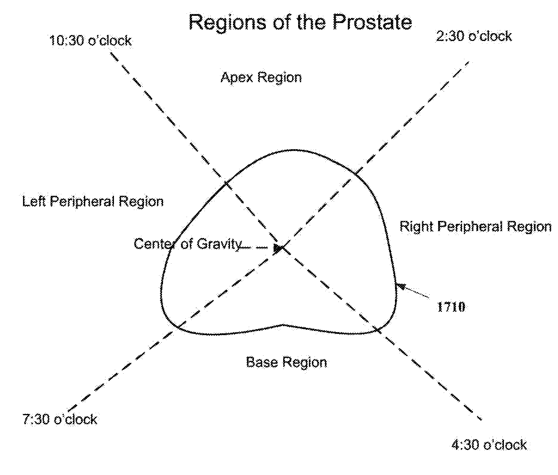
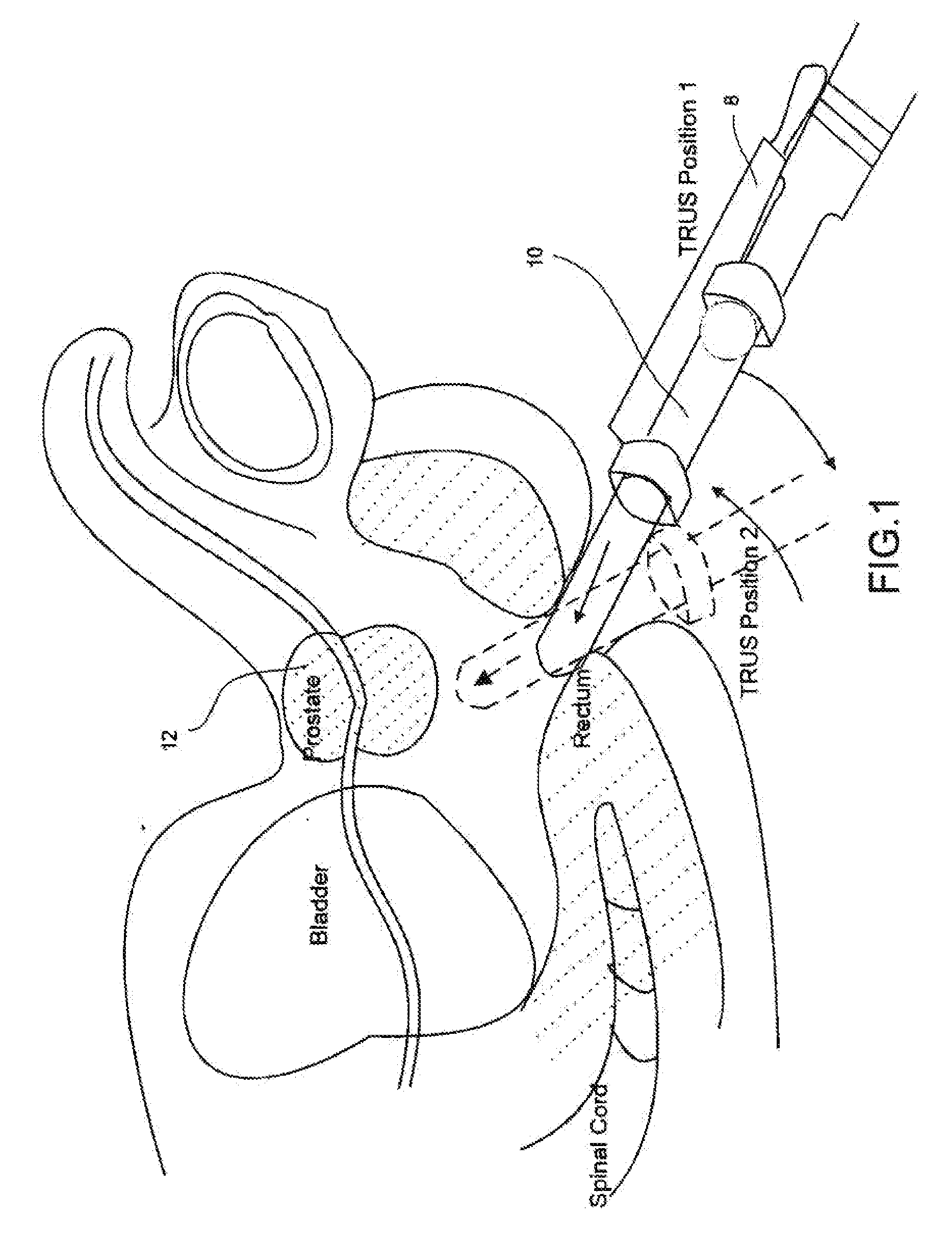
[0043]Reference will now be made to the accompanying drawings, which assist in illustrating the various pertinent features of the present disclosure. Although the present disclosure is described primarily in conjunction with transrectal ultrasound imaging for prostate imaging, it should be expressly understood that aspects of the present invention might be applicable to other medical imaging applications. In this regard, the following description is presented for purposes of illustration and description.

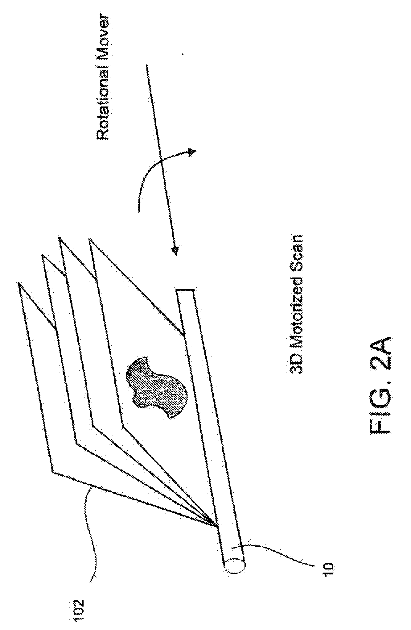
[0044]In the process disclosed herein, a prostate capsule volume computation is made with an automated segmentation method using 2-D ultrasound image. In this process, a 3-D ultrasound prostate image is sliced into the series of contiguous 2-D images, in either a rotational manner, about an axis approximately through the center of the prostate, with a uniform angular spacing (e.g., 3°), or in a parallel manner, with a uniform spacing (e.g. 1 mm). Segmentation for the 2D ultrasound im...


PUM


Abstract

An improved system and method (i.e. utility) for segmentation of medical images is provided. The utility fits an estimated boundary on a structure of interest in an automated selection and lifting process. The estimated boundary may be a model boundary that is generated actual boundaries of like structures. In one arrangement, the boundaries may be selected based on the age and/or ethnicity of a patient. In further arrangements, narrow band processing is performed to estimate the actual boundary of the structure of interest.

Description

CROSS REFERENCE TO RELATED APPLICATION[0001]This application claims priority under 35 U.S.C, §119 to U.S. Provisional Application No. 60 / 908,439 entitled, “AN IMPROVED METHOD OF OBJECT RECOGNITION SYSTEM,” and having a filing date of Mar. 28, 2007, the entire contents of which are incorporated herein by reference.FIELD[0002]The present disclosure pertains to the field of medical imaging, and more particular to the segmentation of medical images to allow for accurate calculation of the volumes of structures of interest within the medical images.BACKGROUND[0003]Medical imaging, including X-ray, magnetic resonance (MR), computed tomography (CT), ultrasound, and various combinations of these techniques are utilized to provide images of internal patient structure for diagnostic purposes as well as for interventional procedures. One application of medical imaging (e.g., 3D imaging) is the measurement of volume and / or assessment of shape of internal structures of interest. These measuremen...

Claims


Application Information

Patent Timeline
Patent Type & Authority Applications(United States)
IPC IPC(8): G06K9/00
CPCG06T7/0083G06T7/0089G06T2207/30081G06T2207/30096G06T2207/20168G06T2207/10132G06T7/149G06T7/12
Inventor SURI, JASJIT S.LI, LU
Owner EIGEN HEALTH SERVICES LLC
Why Eureka
Try Eureka