Method and apparatus to process endoscope images

a technology of endoscope and endoscope body, applied in image data processing, color television, television systems, etc., can solve the problems of unsatisfactory resolution of whitish tissue structures, only inadequate improvement of detection accuracy by conventional image processing techniques, etc., to achieve the effect of eliminating color saturation control effects

Inactive Publication Date: 2008-11-20
OLYMPUS WINTER & IBE
View PDF11 Cites 5 Cited by
  • Summary
  • Abstract
  • Description
  • Claims
  • Application Information

AI Technical Summary

Benefits of technology

[0007]When mapping the color saturation, the mapping function (also “characteristic line”) may be selected in a manner that the zones of higher saturation shall be amplified in order to raise the color saturation contrast compared to the less saturated fasciae or nerves. Preferably however, such a mapping function shall be selected that the color saturation shall be further attenuated in the zones of lesser color saturation. This feature reinforces for instance the fasciae's milky appearance, while strongly colored other organs keep their appearance unchanged. In this manner the image will appear more natural.
[0009]The mapping function is advantageously adjustable to values even applicable to the entire image. Illustratively a mapping function well suited to display certain fasciae may be selected, or a mapping function especially appropriate to display nerves. Also the mapping function may be switched to “Identity”, in order to entirely eliminate color saturation control effects, to allow examining of the natural image.

Problems solved by technology

Whitish tissue structures raise a problem which to-date has not been overcome satisfactorily.
Their detectability is improved only inadequately by conventional image processing techniques.

Method used


Image

  • Method and apparatus to process endoscope images
  • Method and apparatus to process endoscope images

Examples


Embodiment Construction

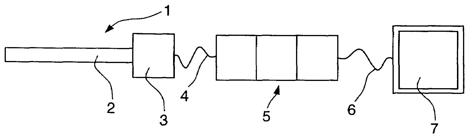
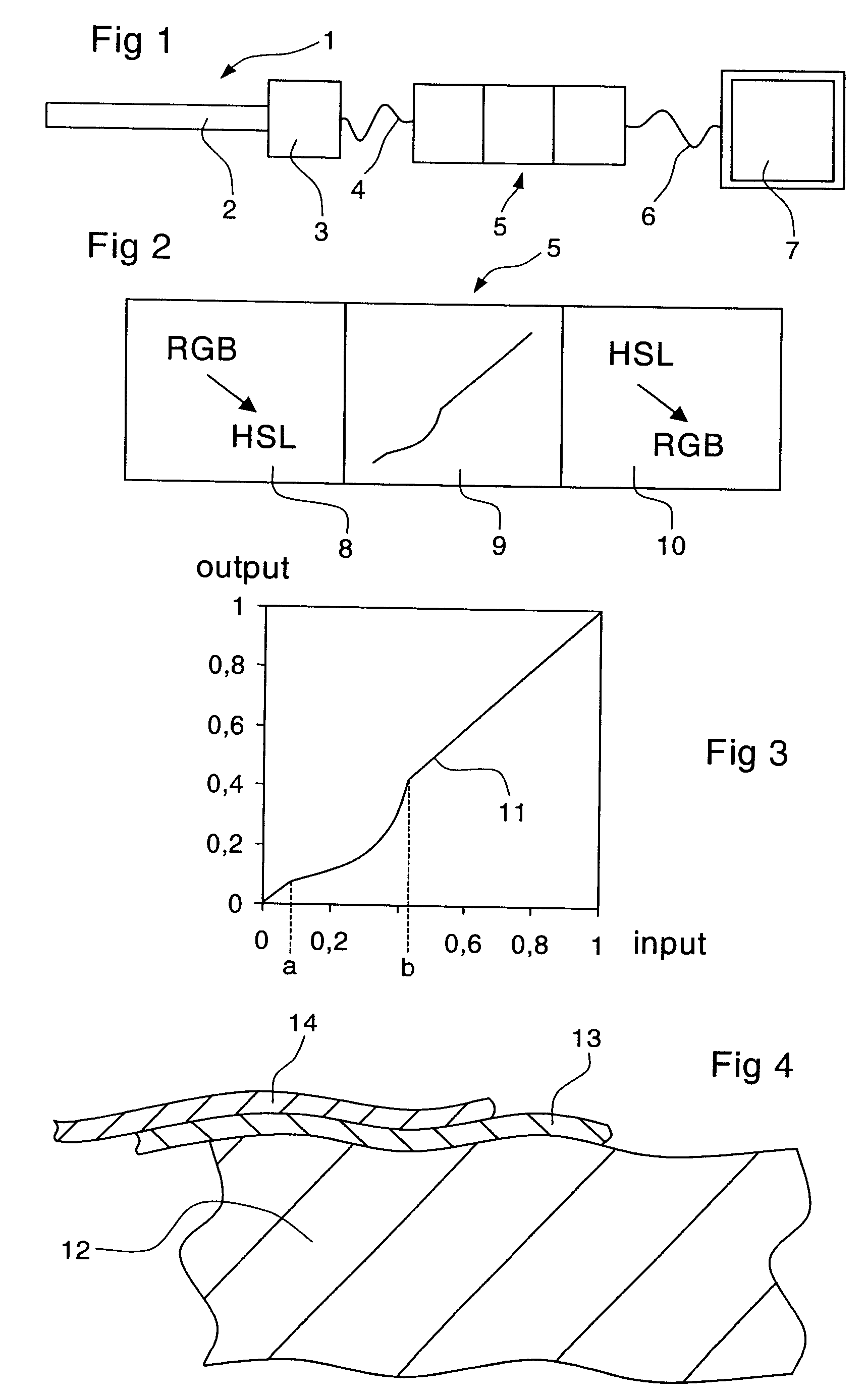
[0018]FIG. 1 shows a medical endoscope 1 fitted with an elongated stem 2, a color video camera 3 being mounted at said stem's proximal end. In another design, the camera 3 also may be configured in the distal end zone of the stem 2 directly behind the camera lens.

[0019]The color video camera 2 is connected to a cable 4 used both for data transmission and illustratively also for electric power, said cable being connected to an image processing unit 5 to which it feeds image data. By means of a cable 6, the image processing unit 5 is connected to an image display unit 7, for instance a conventional monitor.

[0020]Illustratively, the endoscope 1 may be used for laparoscopy and in that case it shall be inserted by its stem 2 through a lancing aperture into the belly area to view organs situated therein. The image seen by the color video camera 3 is recorded and transferred to the image processing unit 5 where it is processed and then displayed on the image display unit 7.

[0021]FIG. 2 sho...


PUM


Abstract

The invention relates to a method improving the images in an image processing unit (5) receiving images from an electronic color video camera (3) of a medical endoscope (1) and then transmitting them to an image display unit (7), and to an endoscope operating on the basis of the said method, where first the pixels of the color components (RGB) are individually mapped into a color space (HSL) wherein the color saturation (S) is independent of the other components (H, L), thereupon the saturation component (S) of each pixel to be mapped is converted by means of a nonlinear mapping function (11) which amplifies the color saturation differential between an upper zone (b->1) and a lower zone (0-b) of the color saturation, lastly the pixels being remapped into an (RGB) color space suitable for image display.

Description

BACKGROUND OF THE INVENTION[0001]Image processing is used in medical endoscopy to gain better insight into definite images.[0002]Known image-processing methods and apparatus are used in medical endoscopy for instance to accentuate given colors, to emphasize the organ structures in weakly structured images, and the like. Conventional image processing procedures are used for such purposes as raising the contrast, color changes and the like.[0003]Whitish tissue structures raise a problem which to-date has not been overcome satisfactorily. Such tissue structures in particular are fasciae, that is thin, transparent skins and also nerves. Such tissues exhibit hardly any color of their own, are translucent and look whitish or milky. The colors of organs underneath them glimmer though them with attenuated intensity.[0004]Whitish tissue structures such as fasciae or nerves—usually situated above organs displaying strongly colored organs, usually can be detected only with difficulty. Their de...

Claims


Application Information

Patent Timeline
Patent Type & Authority Applications(United States)
IPC IPC(8): A61B1/04H04N7/18
CPCA61B1/042G06T5/009G06T5/40G06T2207/10024G06T2207/10068H04N9/67
Inventor AACH, TILSTEHLE, THOMAS
Owner OLYMPUS WINTER & IBE
Why Eureka
Try Eureka