

Coatings and Biomedical Implants Formed From Keratin Biomaterials
a biomaterial and coating technology, applied in the field of keratin biomaterials, can solve the problems of ineffective normal methods used for dissolving proteins with keratin, scientists are confused by the behavior of some purified proteins, and achieve the effect of inhibiting blood coagulation
- Summary
- Abstract
- Description
- Claims
- Application Information
AI Technical Summary
Benefits of technology
Problems solved by technology
Method used
Image

Examples
example 1
Crude Keratose Samples
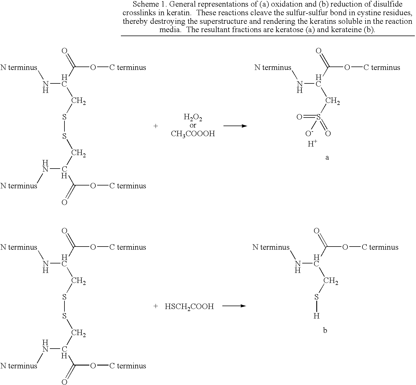
[0091]Keratose fractions were obtained using a method based on that of Alexander and coworkers. However, the method was substantially modified to minimize hydrolysis of peptide bonds. Briefly, 50 grams of clean, dry hair that was collected from a local barber shop was reacted with 1000 mL of an aqueous solution of 2 w / v % peracetic acid (PAA) at room temperature for 12 hr. The oxidized hair was recovered using a 500 micron sieve, rinsed with copious amounts of DI water, and the excess water removed. Keratoses were extracted from the oxidized hair fibers with 1000 mL of 100 mM Trizma® base. After 3 hours, the hair was separated by sieve and the liquid neutralized by dropwise addition of hydrochloric acid (HCl). Additional keratoses were extracted from the remaining hair with two subsequent extractions using 1000 mL of 0.1M Trizma® base and 1000 mL of DI water, respectively. Each time the hair was separated by sieve and the liquid neutralized with HCl. All three ...
example 2
Crude Kerateine Samples
[0092]Kerateine fractions were obtained using a modification of the method described by Goddard and Michaelis. Briefly; the hair was reacted with an aqueous solution of 1M TGA at 37° C. for 24 hours. The pH of the TGA solution had been adjusted to pH 10.2 by dropwise addition of saturated NaOH solution. The extract solution was filtered to remove the reduced hair fibers and retained. Additional keratin was extracted from the fibers by sequential extractions with 1000 mL of 100 mM TGA at pH 10.2 for 24 hours, 1000 mL of 10 mM TGA at pH 10.2 for 24 hours, and DI water at pH 10.2 for 24 hours. After each extraction, the solution was centrifuged, filtered, and added to the dialysis system. Eventually, all the extracts were combined and dialyzed against DI water with a 1 KDa nominal low molecular weight cutoff membrane. The solution was concentrated, titrated to pH 7, and stored at approximately 5% total protein concentration at 4° C. Alternately, the concentrated ...
example 3
Ion Exchange Chromatography
[0093]Just prior to fractionation, keratose samples were re-dissolved in ultrapure water and titrated to pH 6 by addition of dilute HCl solution. Kerateine samples were titrated to pH 6 by careful addition of dilute HCl solution as well. The samples were loaded onto a 200 mL flash chromatography column containing either DEAE-Sepharose (weakly anionic) or Q-Sepharose (strongly anionic) exchange resin (50-100 mesh; Sigma-Aldrich, Milwaukee, Wis.) with gentle pressure and the flow through collected (acidic keratin). A small volume of 10 mM Trizma® base (approximately 200 mL) at pH 6 was used to completely wash through the sample. Basic keratin was eluted from the column with 100 mM tris base plus 2M NaCl at pH 12. Each sample was separately neutralized and dialyzed against DI water using tangential flow dialysis with a LMWCO of 1 KDa, concentrated by rotary evaporation, and freeze dried.
PUM
| Property | Measurement | Unit |
|---|---|---|
| pH | aaaaa | aaaaa |
| pH | aaaaa | aaaaa |
| molecular weights | aaaaa | aaaaa |
Abstract
Description
Claims
Application Information