

Protein markers for the detection of thyroid cancer metastasis
a thyroid cancer and metastasis technology, applied in the field of protein markers for the detection of thyroid cancer metastasis, can solve the problems of not directly identifying the gene expression changes that occur in the metastatic cells, and unable to detect the metastases by current technologies
- Summary
- Abstract
- Description
- Claims
- Application Information
AI Technical Summary
Benefits of technology
Problems solved by technology
Method used
Image

Examples
example i
Materials and Methods
[0040]A. Generation of SAGE libraries. Matched tissues of a normal thyroid (NT), a papillary thyroid carcinoma (PTC) and its lymph node metastasis (LNM) were chosen for SAGE (Case 1, Table 1). This matched set was chosen in part because the sample quality was high, measured by the high percentage of tumor cells observed by H & E histopathology performed on frozen sections from primary and metastatic tumors. The primary sample was from the tumor core, in an attempt to avoid the capsule and surrounding normal tissue. To eliminate the expression of normal lymph node cells without any metastasis a SAGE library was generated from a normal lymph node (NL) (Stratagene La Jolla, Calif., USA). The libraries were constructed using NlaIII as the anchoring enzyme as described in the original SAGE procedure (7) and the ditag containing plasmid inserts were sequenced through the SAGE portion of the Cancer Genome Anatomy Project (8). Tags were extracted from sequence text file...
example ii
Analysis of SAGE Data
[0048]A total of 481,775 SAGE tags were obtained from 4 libraries, representing 160,951 unique transcript tags. The number of SAGE tags per library ranged from 99,911 (NL) to 143,689 (PTC). Tag numbers were normalized to 200,000-tags / library.
[0049]To identify genes potentially involved in metastasis, a comparison between primary tumor and LNM libraries was performed. Monte Carlo simulations yielded 498 tags statistically significant at P value of ≦0.001 or 319 tags at a P value of 0.0001. Thirty-one of the 498 transcripts were highly expressed in the metastasis library and not expressed or expressed at low levels in primary tumor, while 47 transcripts were under-expressed in the metastasis library. To refine our analysis, the transcript expression of the metastasis-associated genes was assessed in the normal lymph node and normal thyroid libraries. Those transcripts with the greatest fold-induction in metastasis library were chosen for validation by qPCR because...
example iii
Relative Levels of Gene Expression for Selected Candidate Genes
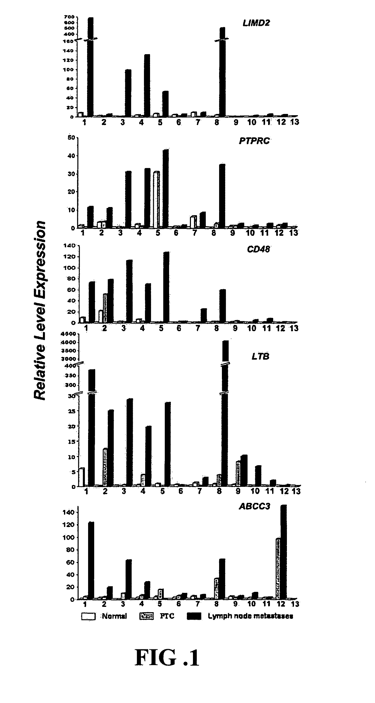
[0051]We used qPCR to test the expression of the transcripts shown in Table 1 in matched sets of normal thyroid, primary PTC and lymph node metastases. This was the first validation set. We compared the results obtained from SAGE with the qPCR results for the samples used to generate NT, PTC and LNM libraries (Case 1, Table 1 and FIG. 1). When the initial samples were used, the difference predicted by SAGE was confirmed for all 11 genes.
[0052]PTPRC was found over-expressed in all metastases analyzed when compared with paired normal thyroid and primary tumor (samples 1-12, FIG. 1) and normal lymph node (sample 13, FIG. 1). LIMD2 and CD48 were consistently expressed in the lymph node-metastases and were not expressed in the normal lymph node (sample 13, FIG. 1) and not expressed or expressed at very low levels in most of matched normal thyroid tissues and / or primary tumors.
[0053]LTB and ABCC3 were remarkably higher in all ...
PUM
| Property | Measurement | Unit |
|---|---|---|
| Tm | aaaaa | aaaaa |
| temperature | aaaaa | aaaaa |
| TG | aaaaa | aaaaa |
Abstract
Description
Claims
Application Information