Shapeable electrosurgical scalpel

a scalpel and electrosurgical technology, applied in the field of shapeable electrosurgical scalpels, can solve the problems of not being able to assist the surgeon in making the smallest cut, the tools provided for the surgeon to remove the tissue are not well suited for performing the procedure, and the surgeon cannot achieve the effect of minimizing the invasiveness of the surgical procedure, and reducing the risk of infection

Inactive Publication Date: 2009-03-26
SENORX
View PDF44 Cites 45 Cited by
  • Summary
  • Abstract
  • Description
  • Claims
  • Application Information

AI Technical Summary

Benefits of technology

[0009]The cutting electrode has a maximum transverse cross-sectional dimension of about 0.007 to about 0.03 inch (0.18-0.76 mm), preferably about 0.008 to about 0.02 inch (0.2-0.5 mm). Elongated cutting electrodes having transverse dimensions of this magnitude may cut large areas of tissue, particularly adipose tissue, with a very effective “clean sweeping motion” with very little pressure against the tissue, thereby creating less trauma to the surgical site and providing for smoother margins of excised tissues. For increased electrode flexibility, the distal section of the cutting electrode may be distally tapered to smaller transverse dimensions. For example, the distal section may taper from a transverse dimension of about 0.01 to about 0.02 inch (0.25-0.51 mm) at the proximal section of the electrode to a smaller transverse dimension of about 0.004 to about 0.01 inch (0.1-25.4 mm) at a distal end of the tapered distal length.
[0015]The invention may also be directed toward a method of performing tissue excision wherein an electrosurgical device is provided having a shapeable elongated electrode with a proximal end that is electrically connected to a high frequency power source and a distal end have a length of exposed cutting surface. The device preferably has a handle configured to hold the electrode and preferably have a mechanism to extend a desired length of exposed electrode out the distal end of the handle for a particular use. The elongated cutting electrode may be preshaped to a desired configuration in its manufacturing process or it may be manually shaped by the physician or other operator just prior to or during the procedure. The cutting electrode is placed in contact with the tissue to be excised and the electrosurgical device is then energized by providing RF power to the cutting electrode from a high frequency power generator. The cutting electrode will readily and smoothly pass through a variety of tissue types including muscular, connective, glandular and fatty tissue. The electrosurgical device may also be energized by the high frequency power generator with wave forms suitable for coagulation of bleeding vessels and tissue. A finger actuated switch on the handle or a dual foot switch situated on the floor allow the user to choose the cutting or the coagulation modes. Other modes may also be provided for other procedures.

Problems solved by technology

While the use of scalpels is widely accepted, they are not designed to minimize the invasiveness of a surgical procedure.
While the practice of removing tissue adjacent to a tissue mass of interest, e.g., a malignant or benign lesion, is followed in many lumpectomy procedures, the tools provided for a surgeon to remove the tissue are not well suited for performing the procedure.
Straight and sculpted blade scalpels do not assist the surgeon in making the smallest cut necessary, and often require the surgeon to essentially dig out the tissue mass.
The damage to the remaining tissues can be significant, resulting in considerable postoperative pain, excessive bleeding, long recovery times, the potential for infection, the potential for depression of the tissues at the surgical site (poor cosmesis) due to the removal of excessive tissue, and surface tissue scarring which is much larger than necessary.
Furthermore, use of these conventional tools and techniques may cause excessive damage to the removed tissue and thus create a tissue specimen having ragged and irregular margins or borders.
This, in turn can lead to inaccurate pathology studies of the excised tissue.
There are some practitioners who believe that a significant number of negative pathology reports (i.e. reports which indicate that the specimen margins are clear of malignant tissue) are false negatives that will most likely result in recurrence of cancer in the patient.
However, such devices typically use small, often pointed active cutting surfaces and the types of devices available to the surgeon who uses electrosurgery are limited.
However, adipose (fatty)) tissue presents a higher impedance to the flow of electrical current than do the other types of tissues, and presents more difficulty in cutting.
These repetitive shallow cuts expose the patient to an increased risk of receiving an unnecessary amount of electrical energy, greater injury to surrounding tissues, greater risk of infection, as well as potentially creating ragged or irregular margins in biopsied tissues.

Method used


Image

  • Shapeable electrosurgical scalpel
  • Shapeable electrosurgical scalpel
  • Shapeable electrosurgical scalpel

Examples


Embodiment Construction

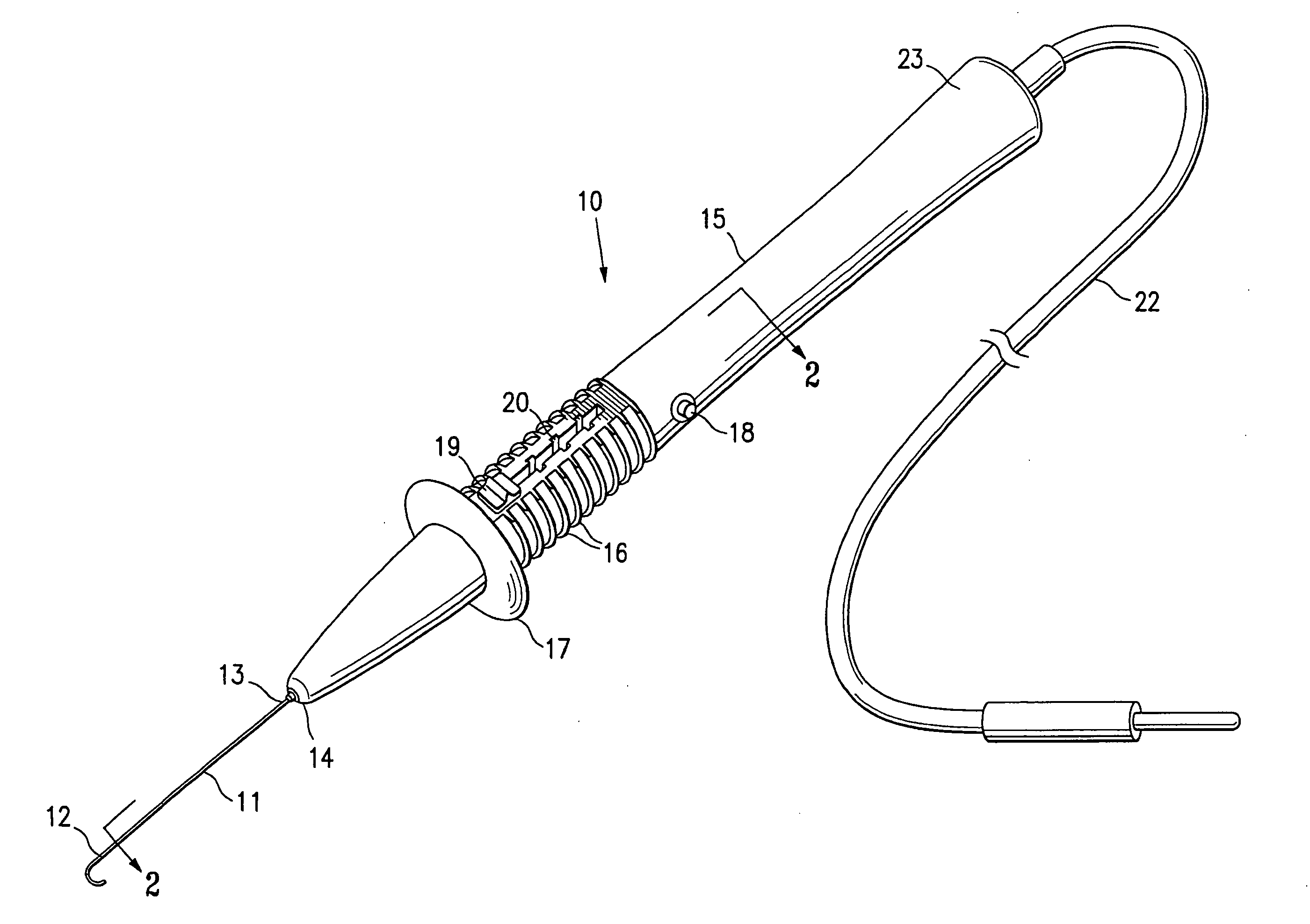
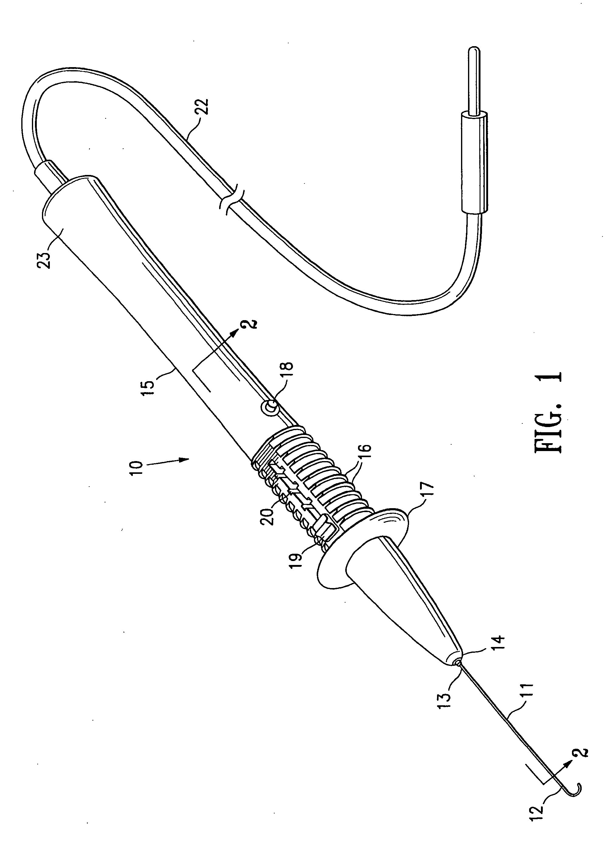
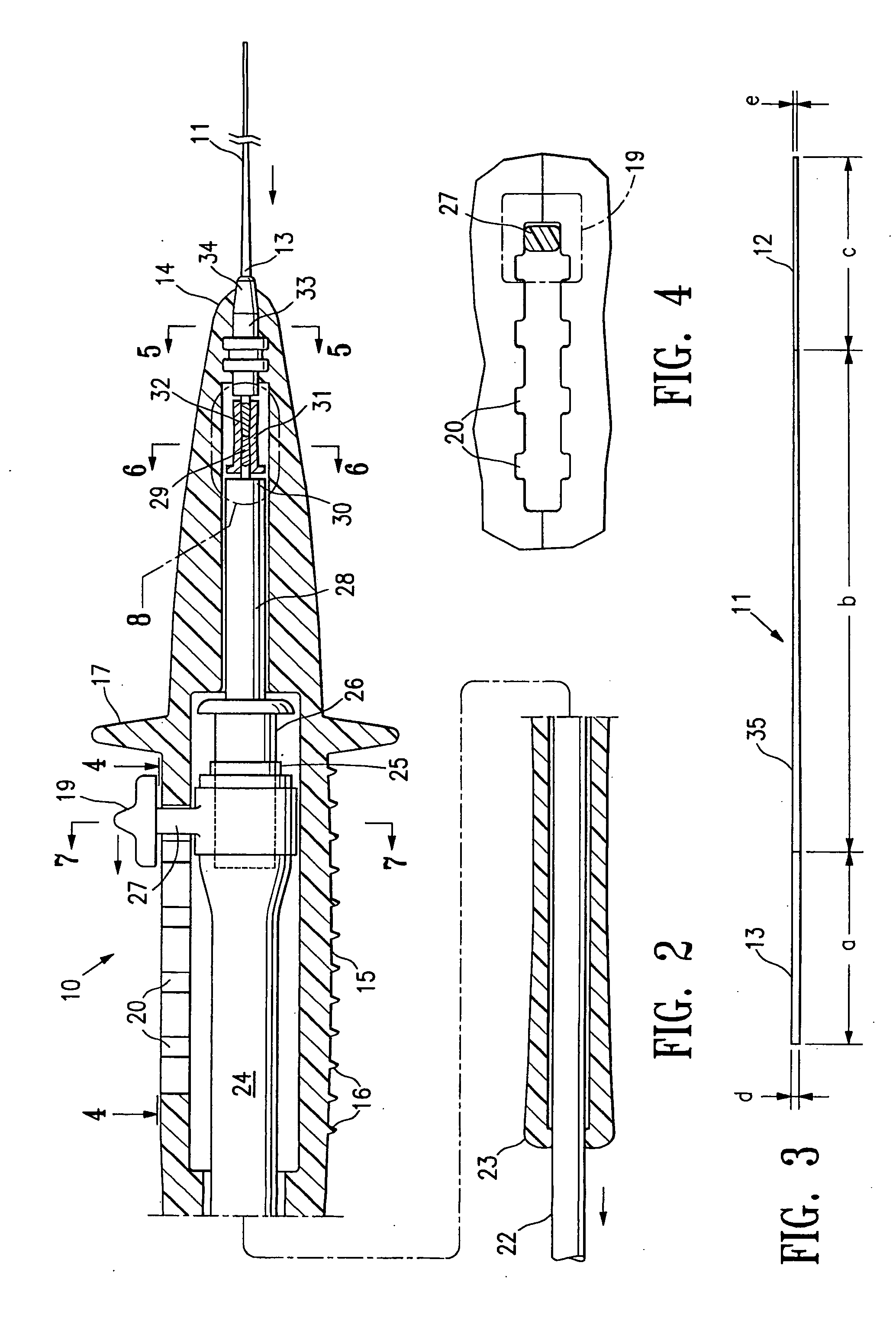
[0036]FIGS. 1-8 depict an electrosurgical cutting device 10 embodying various features of the invention which generally has a cutting electrode 11 with a free or exposed distal portion 12 and a proximal portion 13 which is secured within the distal end 14 of handle 15. The exterior of handle 15 is provided with ridges 16 configured for gripping by the physician or other operator to allow control of the device during operation and a radially extending flange 17 to protect the hand of the operator during operation of the electrosurgical device. The handle 15 is provided with a button type switch 18 for switching an RF electrode power source (not shown) to an active or “on” position or to an inactive or “off” position. A switching function may also be provided for alternative modes such as for coagulation. As best shown in FIGS. 2 and 4, the handle 15 may be provided with a thumb slide 19 to allow axial translation of an electrode assembly within the handle with detents 20 being provid...


PUM


Abstract

The invention is directed to an electrosurgical device having a shapeable elongate cutting electrode having a free distal end with an exposed length of at least 0.5 inch and secured by its proximal end to the distal end of a handle. The electrosurgical device is designed for use with a high frequency electrosurgical generator which has an output at a frequency of between about 1 MHz and about 10 MHz, preferably about 3 to about 8 MHz. Preferably, the output has an essentially sinusoidal waveform with little harmonic distortion. The methods provide for the enhanced cutting of a variety of tissue including muscular, connective, glandular and fatty tissue. The device is particularly suitable in performing a breast biopsy.

Description

RELATED APPLICATIONS[0001]This application is continuation of copending application Ser. No. 10 / 446,507, filed May 27, 2003, which is a continuation of application Ser. No. 09 / 725,265, filed Dec. 29, 2000, which is a continuation-in-part of copending U.S. application Ser. No. 09 / 337,666, filed Jun. 22, 1999, all of which are incorporated herein by reference and from all which priority is claimed.BACKGROUND OF THE INVENTION[0002]Surgical lesion removal has traditionally been performed using a variety of surgical tools and techniques, some of which are specially adapted for a particular procedure. For example, large lesion removal from, e.g., the human breast, is typically attempted through an open incision using an ordinary surgical knife or scalpel. While the use of scalpels is widely accepted, they are not designed to minimize the invasiveness of a surgical procedure. During a surgical procedure, it is usually necessary to form an incision which is much larger than the lesion which...

Claims


Application Information

Patent Timeline
Patent Type & Authority Applications(United States)
IPC IPC(8): A61B18/18A61B18/14A61B10/00A61B10/02A61B17/32
CPCA61B10/0266A61B17/32002A61B18/14A61B18/1402A61B2018/1861A61B2018/00601A61B2018/1405A61B2018/1407A61B2018/00208
Inventor QUICK, RICHARD L.SHABAZ, MARTINKUSSMAN, DANLUBOCK, PAUL
Owner SENORX
Why Eureka
Try Eureka