
Catheter deliverable foot implant and method of delivering the same

a delivery device and catheter technology, applied in the field of subtalar joint and first metatarsalphalangeal implants, can solve the problems of excessive movement in the subtalar joint of the foot, reduce the desirability of the valenti procedure and device, and achieve the effect of minimal invasiveness

Inactive Publication Date: 2009-03-26
CACHIA VICTOR V
View PDF21 Cites 23 Cited by
  • Summary
  • Abstract
  • Description
  • Claims
  • Application Information

AI Technical Summary

Benefits of technology

The solution provides precise alignment and stability with reduced tissue disruption, shorter recuperation times, and improved imaging capabilities, minimizing trauma and postoperative pain while effectively treating subtalar and metatarsal-phalangeal joint disorders.

Problems solved by technology

These changes may eventually result in foot and ankle pain, tendonitis, plantar fasciitis and hallux valgus, hallux limitus and functional disorders of the knees, hips and back.
Although there are several causes of flat feet, one frequent cause is excessive motion in the subtalar joint of the foot.
However, several problems reduce the desirability of the Valenti procedure and device.
Because of its frusto-conical shape and the manner in which it is inserted, the Valenti device is difficult to precisely position in the subtalar joint and difficult to ensure that the proper amount of calcaneal eversion has been eliminated.
Furthermore, it is generally difficult to locate the device properly within the tarsal canal because the implant must be threaded at least 3 to 5 millimeters medial to the most lateral aspect of the posterior facet for correct placement.
Because of its polyethylene construction, the device cannot be imaged using radiography (X-ray) to determine whether the proper position has been achieved.
Another site of frequent foot problems is the first metatarsal-phalangeal joint.
These options are designed to relieve pain and make it easier to walk and engage in physical activities, but they do not address the underlying cause of bunions.
For some patients, non-surgical treatment is sufficient, but surgical intervention is considered if the bunions are progressive or if non-operative treatments provide inadequate improvement.
The most severe and disabling bunions often require extensive joint realignment, reconstruction, implants or joint replacement.
Significant morbidity and recuperation time is required for such procedures.
First MTP-related problems also occur from repetitive trauma to the area and from arthritis.
Over time, active persons can put continuous stress on the first MTP joint that eventually wears out the cartilage and lead to the onset of arthritis.
This condition, known as hallux rigidus, causes loss of movement and pain in the joint.
In most situations, non-operative treatments can be prescribed to provide relief, but those with advanced disease might need surgery, especially when the protective covering of cartilage deteriorates, leaving the joint damaged and with decreased range of motion.
Again, significant morbidity results from these procedures and an extended recovery time is required.

Method used


Image

  • Catheter deliverable foot implant and method of delivering the same
  • Catheter deliverable foot implant and method of delivering the same
  • Catheter deliverable foot implant and method of delivering the same

Examples


Embodiment Construction

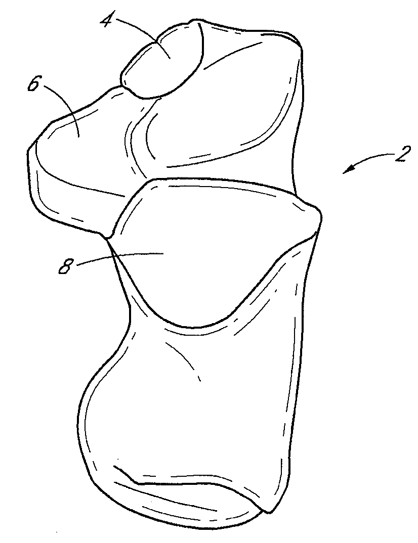
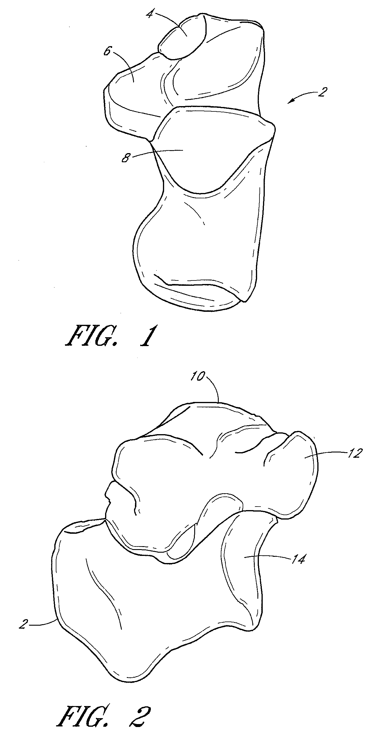
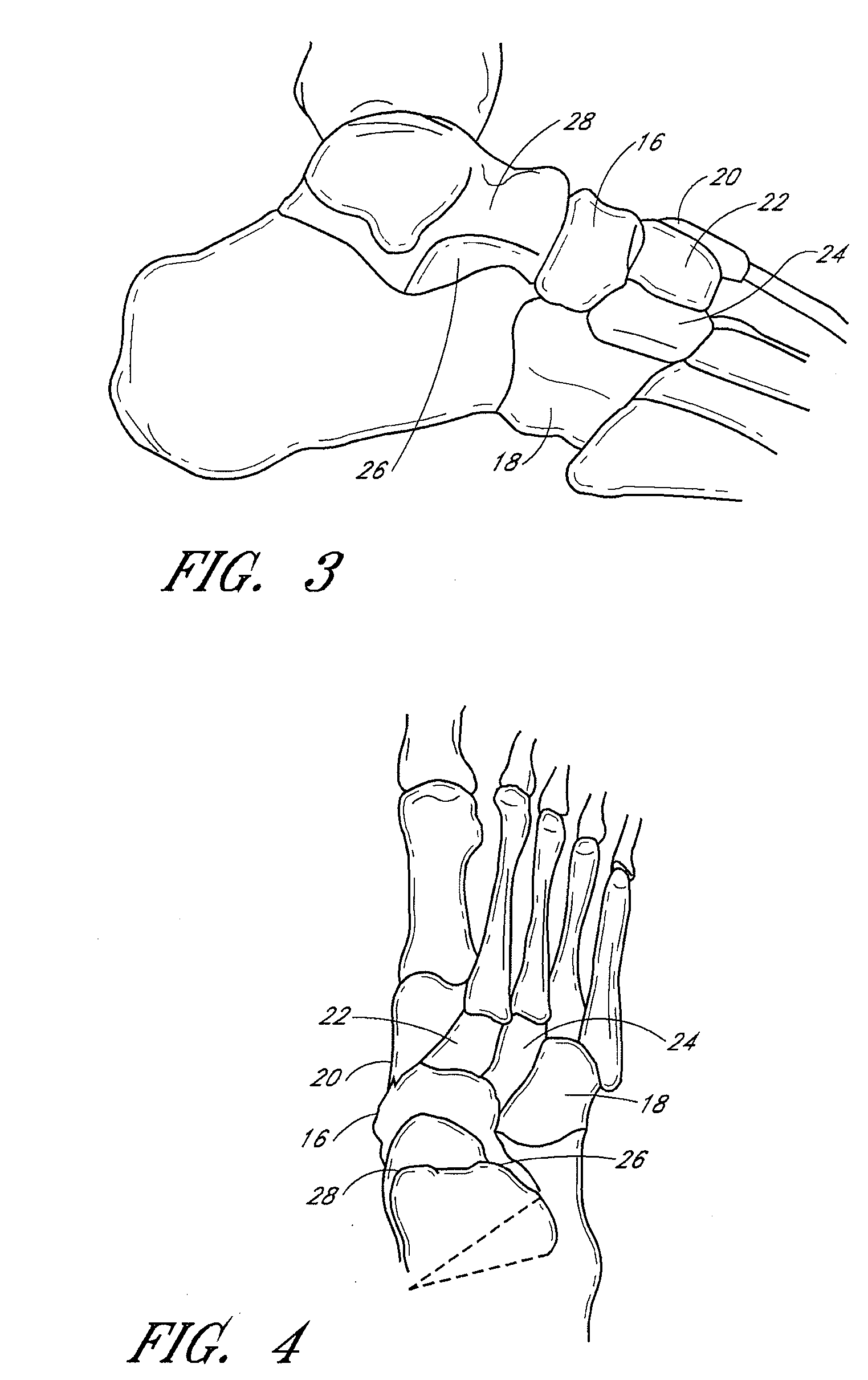
[0056]The talus and calcaneus form the bones of the hindfoot. The talus is a bone with no muscular attachments, but is stabilized by ligaments and cradled by the tendons passing from the leg to the foot. As shown in FIG. 1, the calcaneus 2 articulates with the talus at the calcaneal anterior 4, middle 6 and posterior facets 8. FIG. 2 depicts the relationship between the talus 10 and calcaneus 2 and the talo-calcaneal surfaces 12, 14 that articulate with the midfoot bones. FIGS. 3 and 4 depict the midfoot bones, including the navicular 16, cuboid 18 and cuneiform bones 20, 22, 24. The sinus tarsi 26, also known as the talocalcaneal sulcus, is an extra-articular anatomic space between the inferior neck 28 of the talus 10 and the superior aspect of the distal calcaneus 2. The space continues with the tarsal canal, a funnel or trumpet-shaped space that extends medially to a small opening posterior to the sustentaculum tali. Sinus tarsi 26 is oriented obliquely from a lateral distal open...


PUM


Abstract

Methods and devices are disclosed for manipulating alignment of the foot to treat patients with flat feet, posterior tibial tendon dysfunction and metatarsophalangeal joint dysfunction. An inflatable implant is positioned in or about the sinus tarsi and / or first metatarsal-phalangeal joint of the foot. The implant is insertable by minimally invasive means and inflatable through a catheter or needle. Inflation of the implant alters the range of motion in the subtalar or first metatarsal-phalangeal joint and changes the alignment of the foot.

Description

CROSS-REFERENCE TO RELATED APPLICATIONS[0001]The present application is a continuation of U.S. patent application Ser. No. 11 / 068,675 filed on Mar. 1, 2005, which claims priority under 35 U.S.C. §119(e) to U.S. Provisional Application No. 60 / 549,767 filed on Mar. 3, 2004, the disclosure of which are incorporated by reference herein in its entirety.BACKGROUND OF THE INVENTION[0002]1. Field of the Invention[0003]This invention relates generally to the field of subtalar joint and first metatarsal-phalangeal implants for treating foot conditions including flat feet, adult posterior tibial tendon dysfunction and metatarsophalangeal joint dysfunction.[0004]2. Description of the Related Art[0005]Pes valgo planus, or flat foot, is a common condition where the arch of a foot is weakened and is unable to properly support the weight of the body. With a flat foot, shock absorption is reduced and misalignment of the foot occurs. These changes may eventually result in foot and ankle pain, tendoni...

Claims


Application Information

Patent Timeline
Patent Type & Authority Applications(United States)
IPC IPC(8): A61F2/42A43B13/38A61B17/00A61B17/56A61F2/00A61F2/30A61F2/46
CPCA61B17/562A61B2017/00557A61F2/4606A61F2002/30205A61F2002/30581A61F2002/30841A61B2017/565A61F2002/30904A61F2002/4223A61F2230/0067A61F2002/4627A61F2002/4628A61B17/844A61F2002/30874A61F2002/30873A61F2/30771A61F2/4202A61F2002/3021A61F2002/4629
Inventor CACHIA, VICTOR V.
Owner CACHIA VICTOR V
Learn More