Method for generating digital test objects

a digital test object and image technology, applied in the field of medical image processing, can solve the problems of incomplete and/or inaccurate quality control, limit the scanner time of use for patients, etc., and achieve the effect of reducing control duration and improving accuracy

Active Publication Date: 2009-04-30
QUALIFORMED +1
View PDF2 Cites 2 Cited by
  • Summary
  • Abstract
  • Description
  • Claims
  • Application Information

AI Technical Summary

Benefits of technology

[0017]scanning a physical test object is no more needed to obtain a digital test object, which represents a substantial time saving for the image processing apparatus test procedure and allows to make the scanner available for the patients;
[0019]the DTO generated by the method of the present invention are easily and quickly created, which reduces their cost and hence the image processing apparatus test cost;
[0020]this production cost reduction allows to contemplate a far more comprehensive set of DTO, and therefore to perform thorougher and more comprehensive tests, which eventually enhances the medical image processing apparatus performances;
[0021]the DTO generated by the method of the present invention are more upgradeable than the ones generated by the prior art and thus can better follow the evolutions and regulations in the field of the medical image processing console quality control;
[0022]the perfect management of the DTO production process offered by the present invention allows to know more fully and accurately the DTO and so to perfectly know the expected results of the processings by the image processing console. In the end, the tests performed with the DTO produced according to the present invention are far more accurate;
[0023]since the input DTO produced according to the present invention are perfectly known, test sequences of image processing apparatus can be created by creating expected output DTO and comparing the expected output DTO to the ones produced by the input DTO processing by the image processing apparatus. The test result processing of the image processing apparatus may then be fully automated, which enhances their accuracy and reduces the control durations.

Problems solved by technology

This method has several drawbacks:before each treatment simulation, the scanner has to be reserved to scan the physical test object, which limits the scanner time of use for the patients;the control of the inherent performances of the virtual simulation console is biased due to the fact that the virtual simulation console does not work on perfect images of the physical test object (PTO), but on images obtained and thus already altered by a scanner.
The method currently used only allows to perform a quality control on the scanner & virtual simulation assembly, not on the virtual simulation assembly alone;the time required for the quality control comprising the TPO image acquisition sequence by the scanner and then the processing by the virtual simulation console is lengthened;finally, taking into account the machining complexity of the materials they involved, the PTO are manually limited and / or adjustable, which leads to incomplete and / or inaccurate quality controls.

Method used


Image

  • Method for generating digital test objects
  • Method for generating digital test objects
  • Method for generating digital test objects

Examples


Embodiment Construction

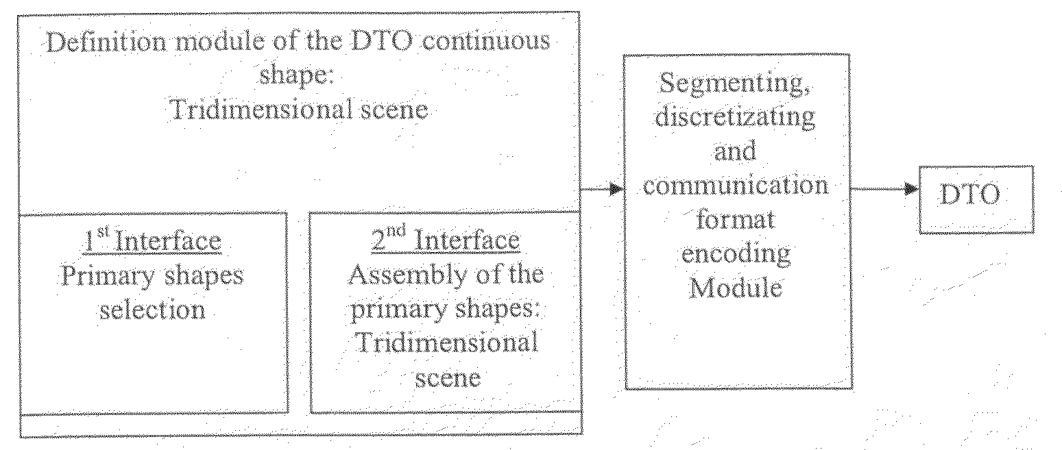
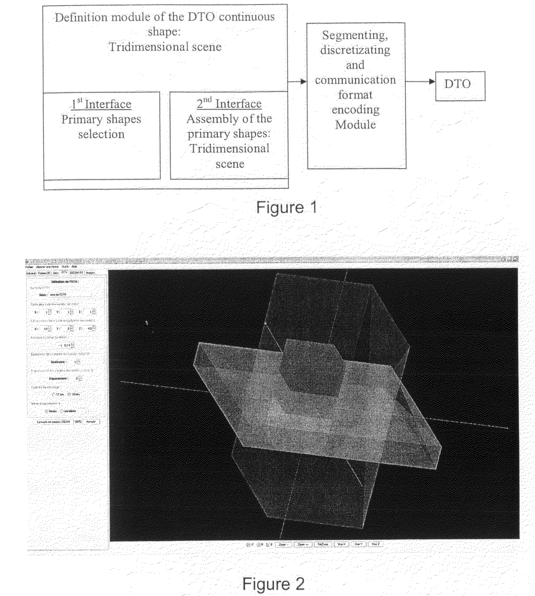
[0051]As can be seen on FIG. 1, the software carrying out the digital test object generation method comprises two main modules.

[0052]The first module is a module that allows to define the tridimensional continuous shape of the digital test object from basic primary shapes. The second module allows to discretize and segment the obtained tridimensional continuous shape into volume elements (voxels) into bidimensional cross-sections. Finally, this second module encodes the bidimensional cross-sections into a format readable by medical imaging apparatus, for example into DICOM format.

[0053]The first module for defining the DTO continuous shape from basic primary shapes contains two distinct interfaces.

[0054]The first interface, the “shape” interface, allows to individually define each basic primary shape, and possibly apply translations and rotations to these basic primary shapes.

[0055]The basic primary shapes can be, for example:[0056]a point, which is then a voxel the center of which ...


PUM


Abstract

The object of the present invention is a computer-implemented method for generating digital images encoded into a format readable by medical imaging apparatus, characterized in that it comprises the steps of:defining, in a tridimensional continuous space, a tridimensional scene comprising a tridimensional continuous shape and an encompassing medium, said tridimensional shape being constituted from basic primary shapes by rotation, translation, overlapping or gathering of one or several basic primary shapes;discretizing the tridimensional scene obtained at the previous step into volume elements (voxels) using a weighting mechanism;segmenting the discretized tridimensional scene into voxels by creating bidimensional cross-sections of the tridimensional scene; andencoding the information describing the obtained bidimensional cross-sections of the tridimensional scene into a format readable by medical imaging apparatus.

Description

CROSS-REFERENCE TO RELATED APPLICATIONS[0001]Applicant claims priority under 35 U.S.C. 119 of French patent application No. 0758612 filed on Oct. 26, 2007.BACKGROUND OF THE INVENTION[0002]The present invention relates to the field of medical image processing, and more particularly to a method for generating digital images in a format readable by medical image processing apparatus, to digital images generated by such a method as well as to a method for the quality control of the medical image processing consoles based on the generated digital images.[0003]Medical image processing consoles are used in many applications, either for a diagnostic purpose, or for a therapeutic purpose, to prepare or perform some treatments, in particular in the radiotherapy field which consists in sterilizing the cancerous tumours by means of high doses of ionizing radiation usually delivered with multiple concentric X-ray beams produced by an linear electron accelerator. Taking into account the potential...

Claims


Application Information

Patent Timeline
Patent Type & Authority Applications(United States)
IPC IPC(8): G06K9/00G06T17/00G06T15/08
CPCG06T15/08G06T2219/2012G06T19/00
Inventor DENIS, ELOISEBEAUMONT, STEPHANEGUEDON, JEAN-PIERRENORMAND, NICOLAS
Owner QUALIFORMED
Why Eureka
Try Eureka