Color mapped magnetic resonance imaging

Inactive Publication Date: 2009-06-04
AURORA IMAGING TECH
View PDF16 Cites 14 Cited by
  • Summary
  • Abstract
  • Description
  • Claims
  • Application Information

AI Technical Summary

Benefits of technology

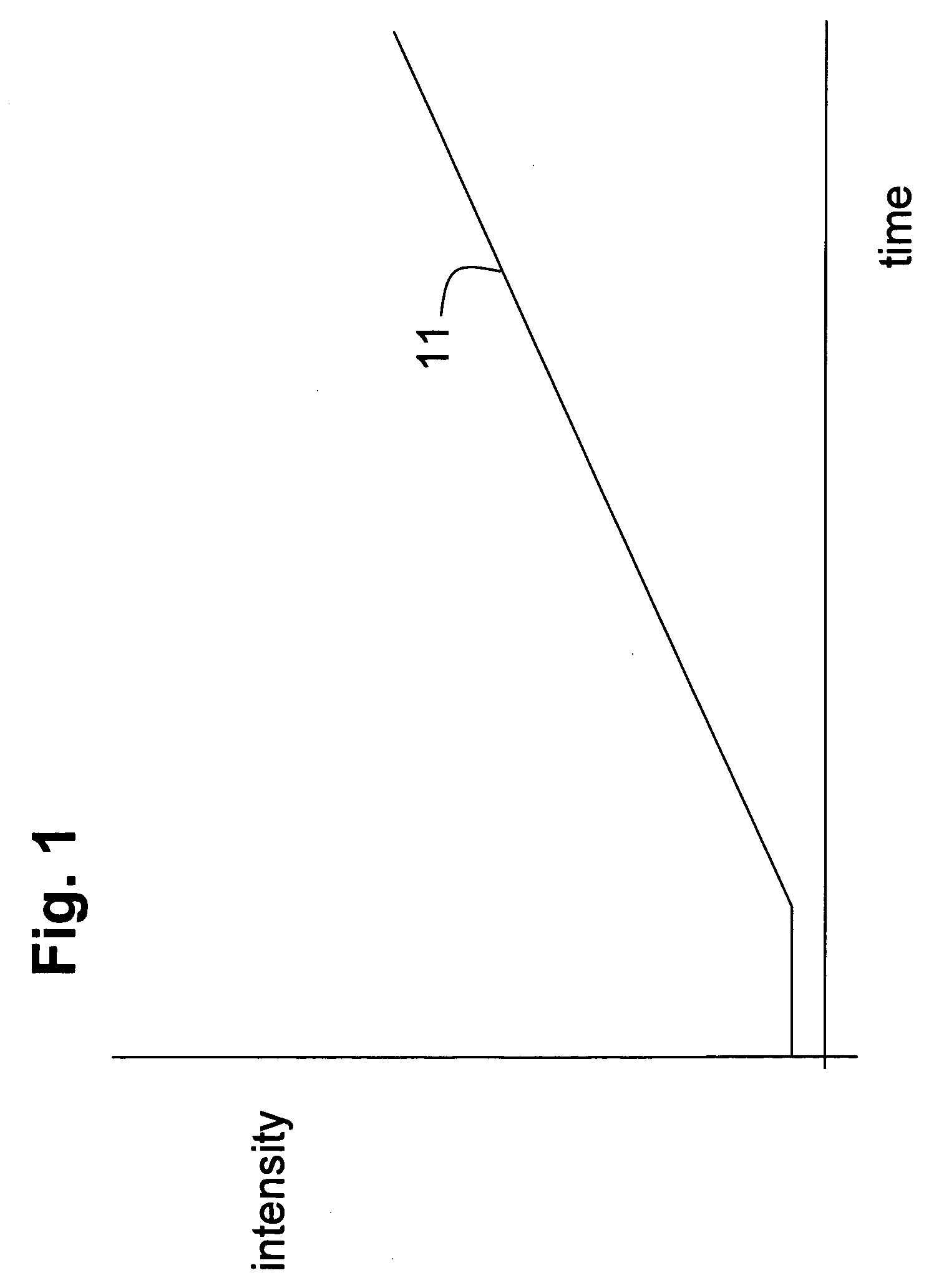
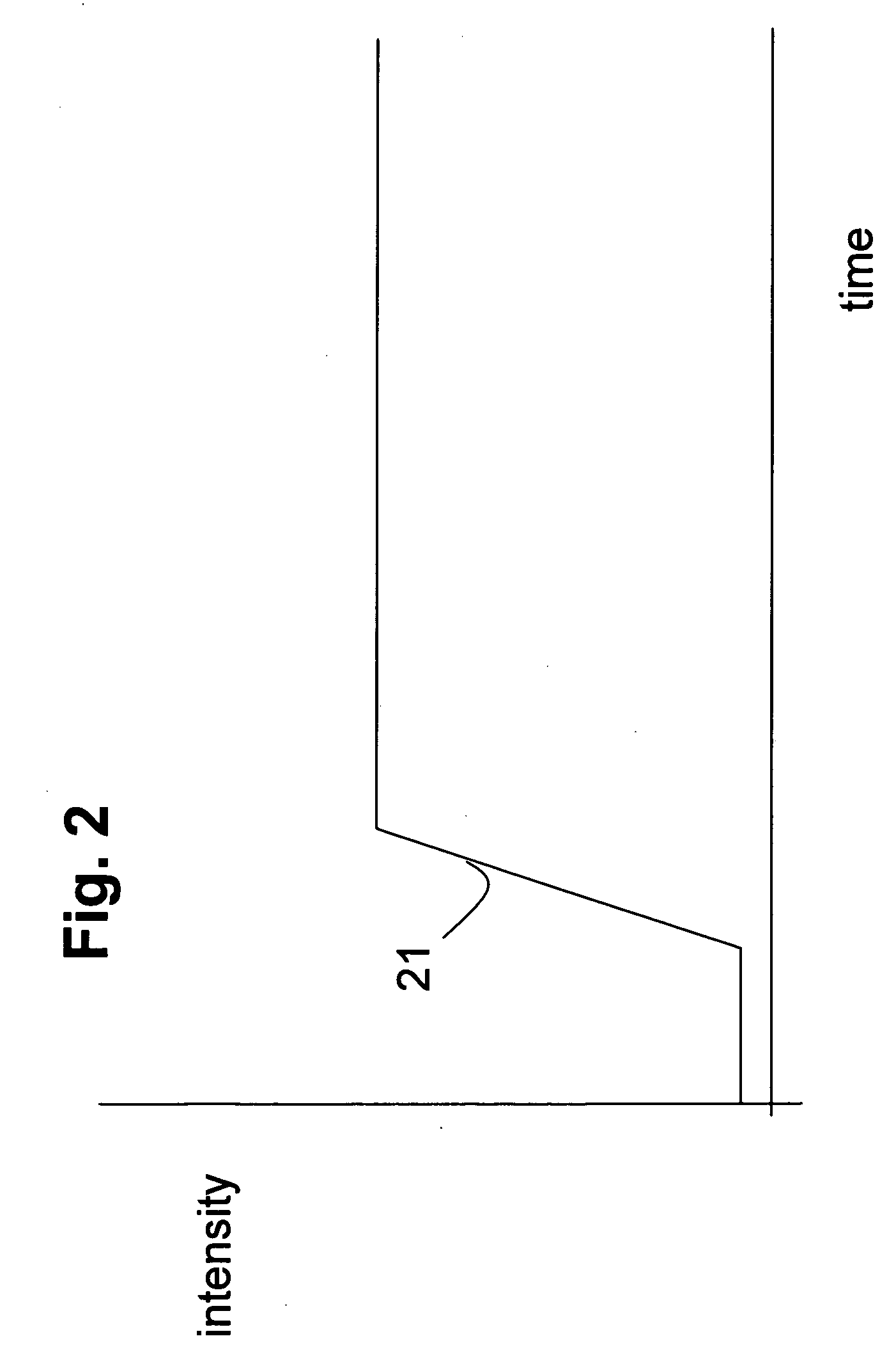
[0024]The invention is advantageous for meaningful display of MRI data with respect to risk, and in particular risk of malignancy for identified points, tissue structures or regions. The system and method also may be used in other ways, for example for parametric color mapping to identify silicone in implants or in leakage from implants, which can be displayed in a distinct predetermined color. To recognize silicone, a silicone-suppressed image can be subtracted from a non-silicone suppressed image and the resultant image can be color coded for silicone in regions that show a detectable presence or a given concentration of silicone. Risk of malignancy can also be assessed in part using precontrast images, especially to improve the contrast associated with some features by subtracting away from the post contrast data at least part of the precontrast image data so as to remove the common mode background image from the displayed characteristics. It is also possible to use a reference image wherein the spatial resolution or T1 and T2 characteristics enable discrimination for compositions such as silicone, in which case subtraction may not be needed. Also, information obtained from subtraction or from a reference image can be supplemented by curve shape information.

Problems solved by technology

Generally, alarm or hot colors include red, magenta, orange, yellow, etc., and are associated with higher levels of risk.

Method used


Image

  • Color mapped magnetic resonance imaging
  • Color mapped magnetic resonance imaging
  • Color mapped magnetic resonance imaging

Examples


Embodiment Construction

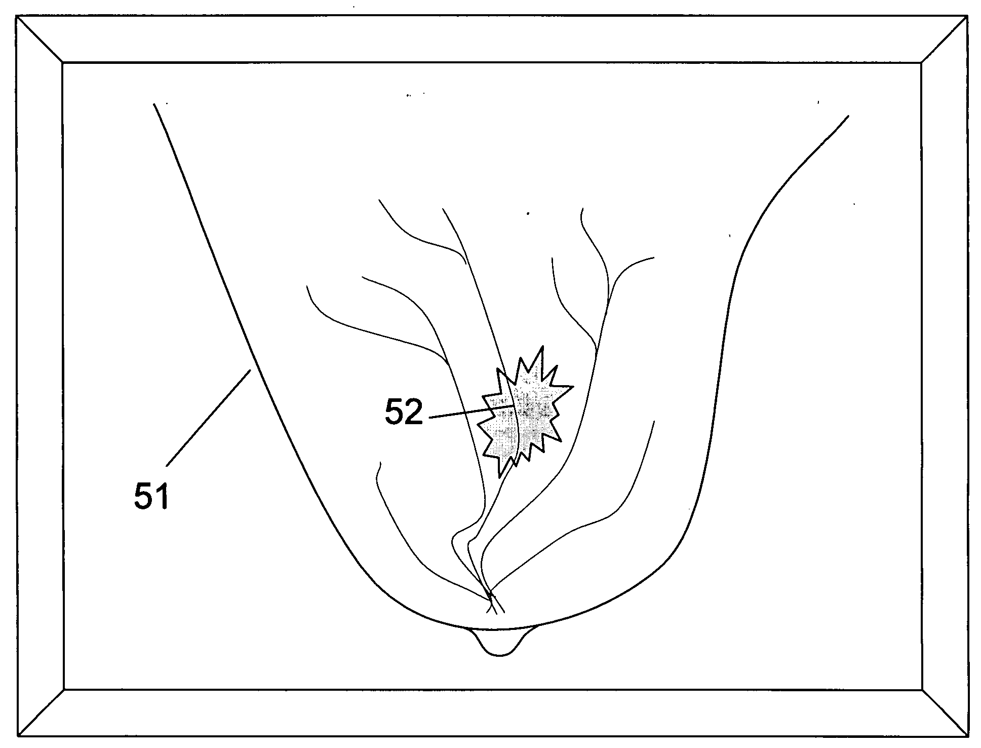
[0031]According to an inventive aspect, imaging data characteristics obtained by nuclear magnetic resonance imaging (MRI) techniques, especially the time-changing effects of perfused contrast agents on the intensity of voxel points in imaged breast tissues. The data are processed to evaluate the risk associated with a given object within the breast according to one or more diagnostic standards that are preferably automated and accomplished in image data processing routines. The processed level of risk perceived for respective voxel points, tissue structures and / or imaged regions, is mapped to the display of intensity as used for visualizing the tissue structures.

[0032]In one embodiment, the amplitude of the MRI response is mapped to the intensity of the image of the corresponding tissues structures. The associated level of risk for the tissue structures or distinct areas thereof, is mapped to a distinct image attribute associated with the displayed image of that tissue structure or ...


PUM


Abstract

A method and system estimate the risk of malignancy of a given region of interest using noninvasive MRI techniques. The determination of risk is based on the morphology and kinetic enhancement of a region of interest. In addition, the method and system use the type of the enhancement curve to determine the level of risk associated with a given region of interest. The region of interest can be a lesion, tumor, or other unknown. The imaging can be done with the aid of a contrast agent. Regions meeting component concentration criteria, time-change dynamic criteria and the like are distinctly colored and displayed on a single image. Different colors can be shown locally to identify predetermined levels of risk, and / or associated with predetermined compositions such as contrast agents, or to show the presence of silicone.

Description

FIELD OF THE INVENTION[0001]The invention relates to techniques for processing and displaying the results of clinical imaging, particularly nuclear magnetic resonance imaging of the human breast for breast cancer screening, diagnosis and / or treatment.BACKGROUND[0002]Lesion morphology and kinetic enhanced contrast analyses can provide information that may enable a skilled practitioner to assess the relative probability that a tissue anomaly or lesion might be malignant when seen in the nuclear magnetic resonance image (MRI) of a breast or other tissue. That is to say, an experienced practitioner or radiology technologist can learn to assess risk based on visual clues that are found in MRI images. There are various different ways that MRI images are obtained, weighted, enhanced by use of perfused agents or by image processing routines and otherwise produced. Thus, it is not clear how best to collect MRI data and to compare different collected images, to optimize the probability that d...

Claims


Application Information

Patent Timeline
IPC IPC(8): A61B5/055
CPCA61B5/055G06T11/001A61B5/7275G16H50/30G16H50/20G16H30/40G16H20/17
Inventor HARMS, STEVEN E.SPANGENBERG, SCOTTHONG, XIAOLE
Owner AURORA IMAGING TECH
Why Eureka
Try Eureka