
Device and method for orthopedic fracture fixation

a technology for fixing and stabilizing devices and fractures, applied in the field of orthopedic fracture fixation and stabilization devices, can solve the problems of severe limitation of activity, constant pain, serious reduction of quality of life, etc., and achieve the effect of preventing the subsequent post-operative loosening of bone screws

Inactive Publication Date: 2009-06-25
INTEGRAL SPINE SOLUTIONS
View PDF15 Cites 47 Cited by
  • Summary
  • Abstract
  • Description
  • Claims
  • Application Information

AI Technical Summary

Benefits of technology

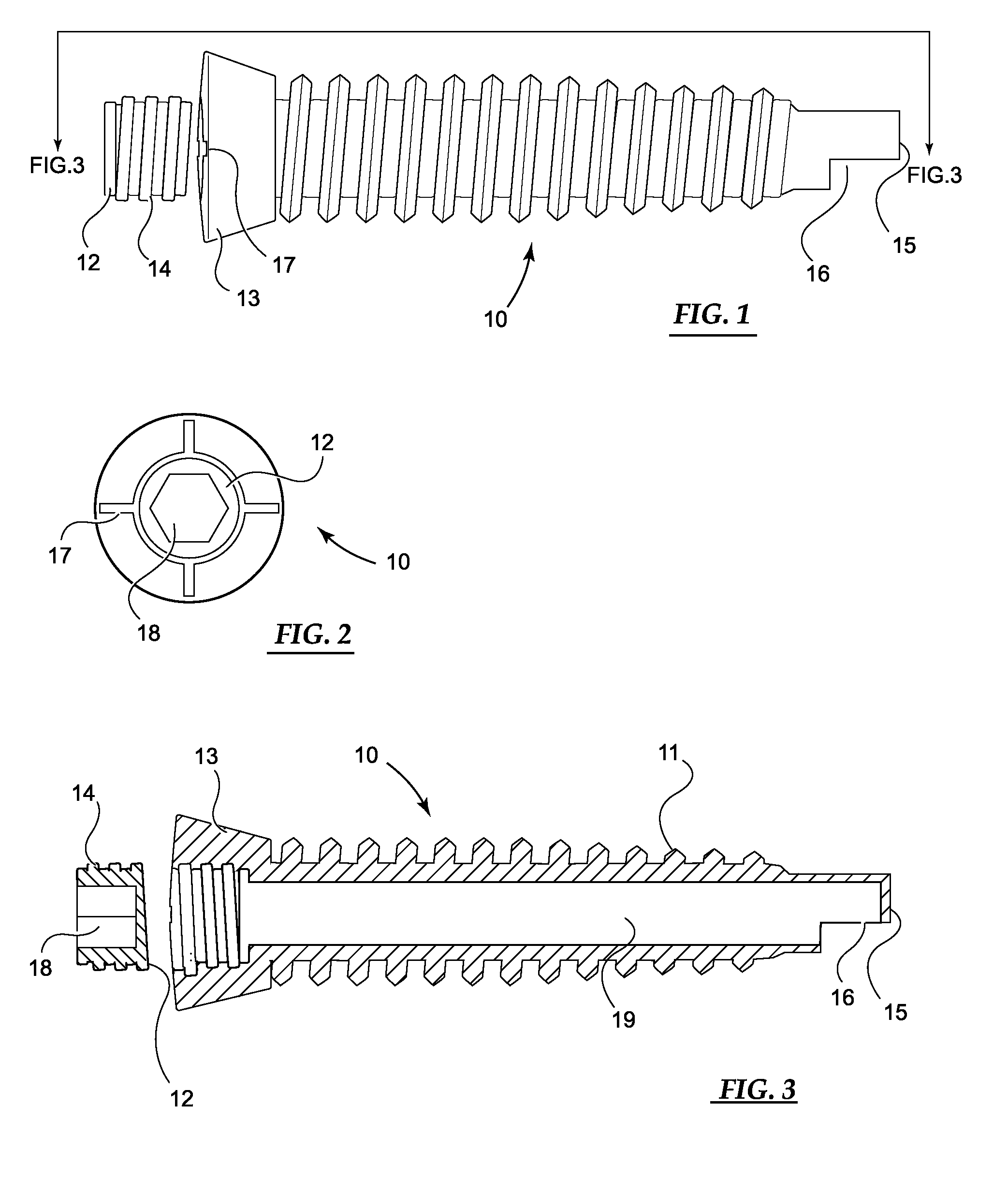
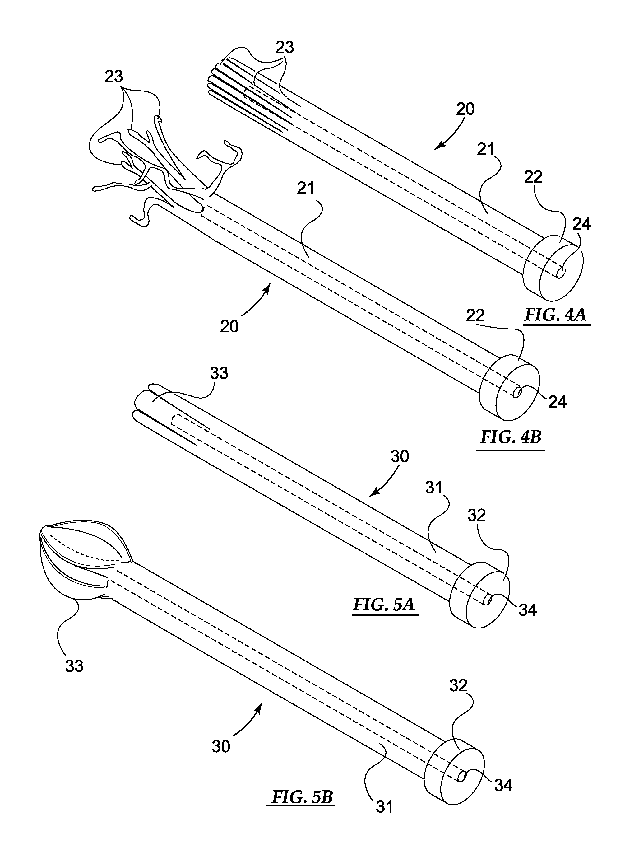
[0024]The present invention provides a reliable integrated system for fracture fixation and other such procedures wherein the delivery of mechanical implants and introduction of flowable compositions such as bone cement into an intraosseous space are integrated. The devices and methods are useful anywhere in the axial or peripheral skeleton of a mammalian body. The systems comprise cannulated orthopedic screws and mechanical expandable implants introducible through the lumen of such screws wherein the system is configured such that the expandable proximal end of the implant remains attached to the cannulated screw to provide an anchor for the implant. In preferred embodiments the cannulated implants are expandable within the intraosseous space to introduce a scaffold-like structure such as branched, basket, mesh umbrella and the like and wherein after subsequent introduction of a flowable composition such as bone cement the device functions to secure the bone screw and prevents the subsequent post-operative loosening of bone screws often observed months or years after the fixation procedure. The devices are also particularly useful for delivering flowable restorative compositions into an osseous cavity.
[0025]A typical embodiment of an orthopedic fracture fixation device of the present invention comprises a cannulated orthopedic screw comprising a through-bore along the longitudinal axis that defines an orthopedic screw lumen extending between an open orthopedic screw proximal end and an open orthopedic screw distal end; a screw cap attachable to the orthopedic screw proximal end that functions to seal the orthopedic screw proximal end; and a cannulated implant sized and configured to be slidably disposable within the through-bore of the orthopedic screw wherein the cannulated implant distal end comprises an expandable region that is disposed beyond the orthopedic screw distal end when the cannulated implant proximal end is disposed completely within the orthopedic screw through-bore and wherein the expandable region of the cannulated implant is expanded to assume a predetermined configuration when disposed within a targeted location in a mammalian body. The combination of the implanted structure and the bone cement stabilize the fractured bone.
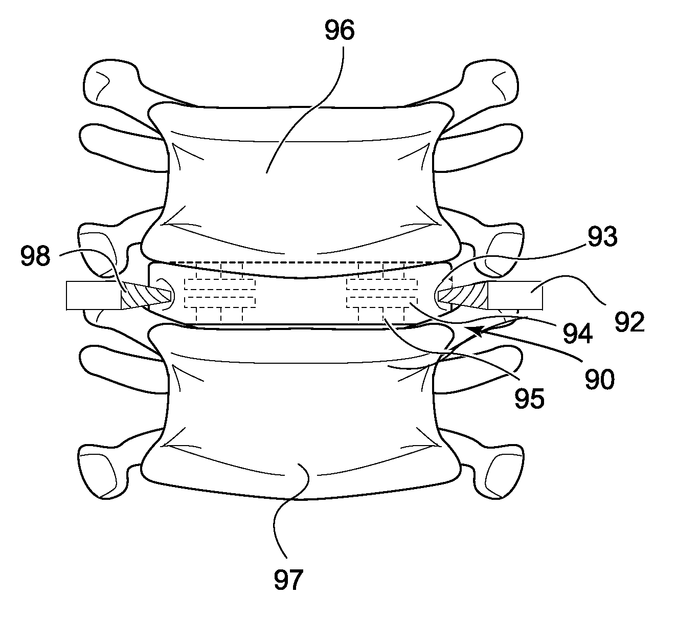
[0026]This devices of the present invention are useful for treatment and stabilization of malignant or traumatic vertebral compression fracture of the spine and the combination of the nitinol implanted structure and the bone cement stabilizes and reconstructs the affected vertebra.
[0027]Certain embodiments of the present invention employ cannulated screws with side ports or holes wherein a pre-shaped expandable implant in a compressed state is inserted inside the cannulated screw and branch-like elements of the implant situated along the shaft of the implant are extend outside of the screw through the sides ports for additional stabilization. Such devices are particularly useful in spinal procedures or in treatment of fractures of long bones like tibia wherein it is desirable to prevent loosening of the screws.

Problems solved by technology

For many people with osteoporosis a spinal fracture results in severely limited activity, constant pain and serious reduction in quality of life.
Additionally, younger individuals may also suffer such vertebral compression fractures, particularly individuals whose bones have become fragile due to the long-term use of steroids or other drugs to treat a variety of diseases such as lupus, asthma and rheumatoid arthritis.
While various types of back bracing devices can also be used, such devices may actually cause weakening of the bone and predispose patients to further fractures in the future.
Although successful vertebroplasty has been shown to alleviate the pain caused by a compression fracture, these techniques are often deficient providing adequate stability and physiologic reconstruction of the spinal column and often prevents patients from return to a previous level of physical activity.
However, as in the vertebroplasty procedures, balloon kyphoplasty techniques are often insufficient in providing adequate stability and physiologic reconstruction of the spinal column and often prevents patients from return to a previous level of physical activity.
While the above referenced devices address problems relating to the viscosity of flowable compositions such as bone cements, they do not address issues relating to the precise control of placement and distribution of such compositions at a targeted injection site.
However, the devices described in these patents deliver the injected material only through the tip of cannula and therefore offer no control of the direction or distribution of the injected material within the organism.
Furthermore, these patents do not teach methods for delivery of restorative material by percutaneous vertebroplasty by which multiple doses of material can be injected.
However, such a cannula with a single opening in the cannula wall as described does not provide sufficient control of cement placement nor degree of directional control required to prevent extravasation in these delicate procedures.
Such a cannula is not suitable for the injection of viscous flowable materials such as bone cement and, since the narrow openings are confined near the tip of the cannula, such a design offers no control over the placement of the fluid.

Method used


Image

  • Device and method for orthopedic fracture fixation
  • Device and method for orthopedic fracture fixation
  • Device and method for orthopedic fracture fixation

Examples


Embodiment Construction

[0045]For the purposes of the invention described in this application, the certain terms shall be interpreted as shown below.

[0046]The term ‘cannulated’ describes the property of an object as being hollow or tubular and affording a passage through its interior length for a flowable materials and suitably sized solid objects such as catheters, rods, wires, threads, and the like.

[0047]The term ‘side-port’ describes any orifice in the wall of a tube, pipe or cannula that allows communication between the lumen of the tube, pipe or cannula and an outside area.

[0048]The term ‘flowable material’ describes any injectable material that flows as a uniform mass when an appropriate pressure is applied. Such flowable materials may comprise solutions, emulsions, suspensions, slurries, pastes, gels, polymerizable monomers, liquid polymers, oligomers, and all mixtures or combinations thereof.

[0049]Basic embodiments of the orthopedic fixation devices of the present invention comprise: a cannulated o...


PUM


Abstract

The present invention relates invention relates to devices and methods for the fixation and stabilization of orthopedic fractures and more specifically to stabilization of compression fractures of spinal vertebrae. The systems comprise cannulated orthopedic screws and mechanical expandable implants introducible through the lumen of such screws. In certain embodiments the cannulated implants are expandable within an intraosseous space to introduce scaffold-like structures such that subsequent introduction of a bone cement forms a composite in situ. The devices and methods are also particularly useful for stabilizing the orthopedic screws.

Description

RELATED APPLICATION DATA[0001]This application claims priority under 35 U.S.C. §119(e) to U.S. Provisional Application Ser. No. 61 / 014,748, filed Dec. 19, 2007.FIELD OF THE INVENTION[0002]This invention relates to devices and methods for the fixation and stabilization of orthopedic fractures. More specifically, these devices and methods apply to stabilization of compression fractures of spinal vertebrae.BACKGROUND[0003]The mammalian spine consists of bones called vertebrae, which are separated by soft cushions referred to as intervertebral discs. The thick portion of bone at the front of each vertebra is referred to as the vertebral body. When a vertebral body collapses, a vertebral compression fracture (VCF) of the bone results. Most vertebral compression fractures are caused by osteoporosis, a disease that causes bones to become brittle and to break easily. Because osteoporosis usually progresses without obvious symptoms, an individual may not be aware that he or she has the disea...

Claims


Application Information

Patent Timeline
Patent Type & Authority Applications(United States)
IPC IPC(8): A61F2/44A61B17/04
CPCA61B17/7001A61F2310/00023A61B17/864A61B2017/00867A61F2/442A61F2002/30331A61F2002/30484A61F2002/30579A61F2002/30598A61F2002/30604A61F2002/30736A61F2002/30784A61F2002/30841A61F2002/448A61F2310/00017A61B17/7098A61F2002/30594
Inventor GEORGY, BASSEMGHOBRIAL, EMAN K. R.
Owner INTEGRAL SPINE SOLUTIONS
Learn More