

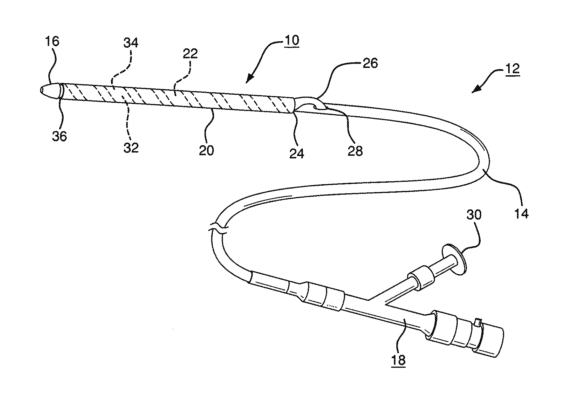
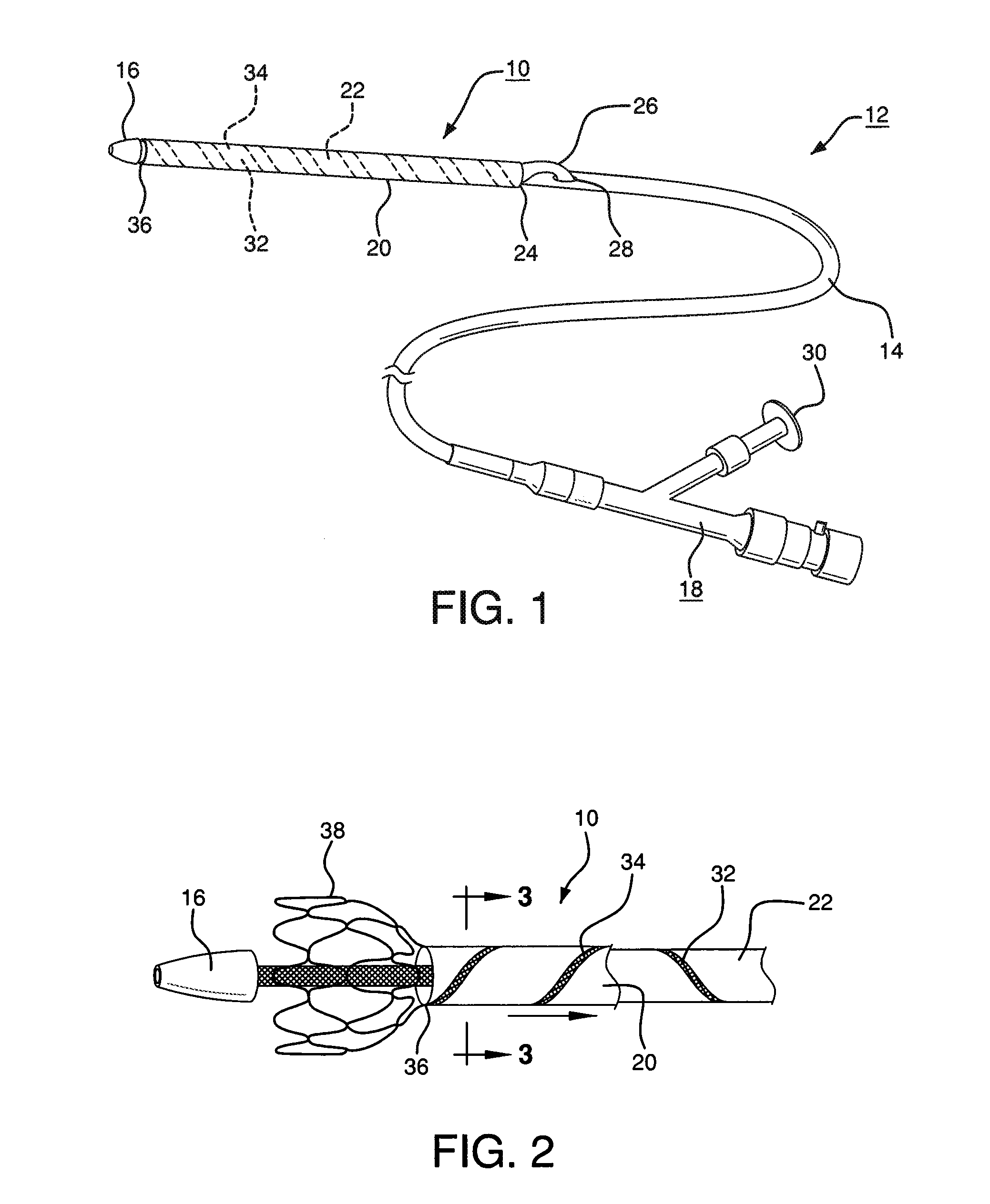
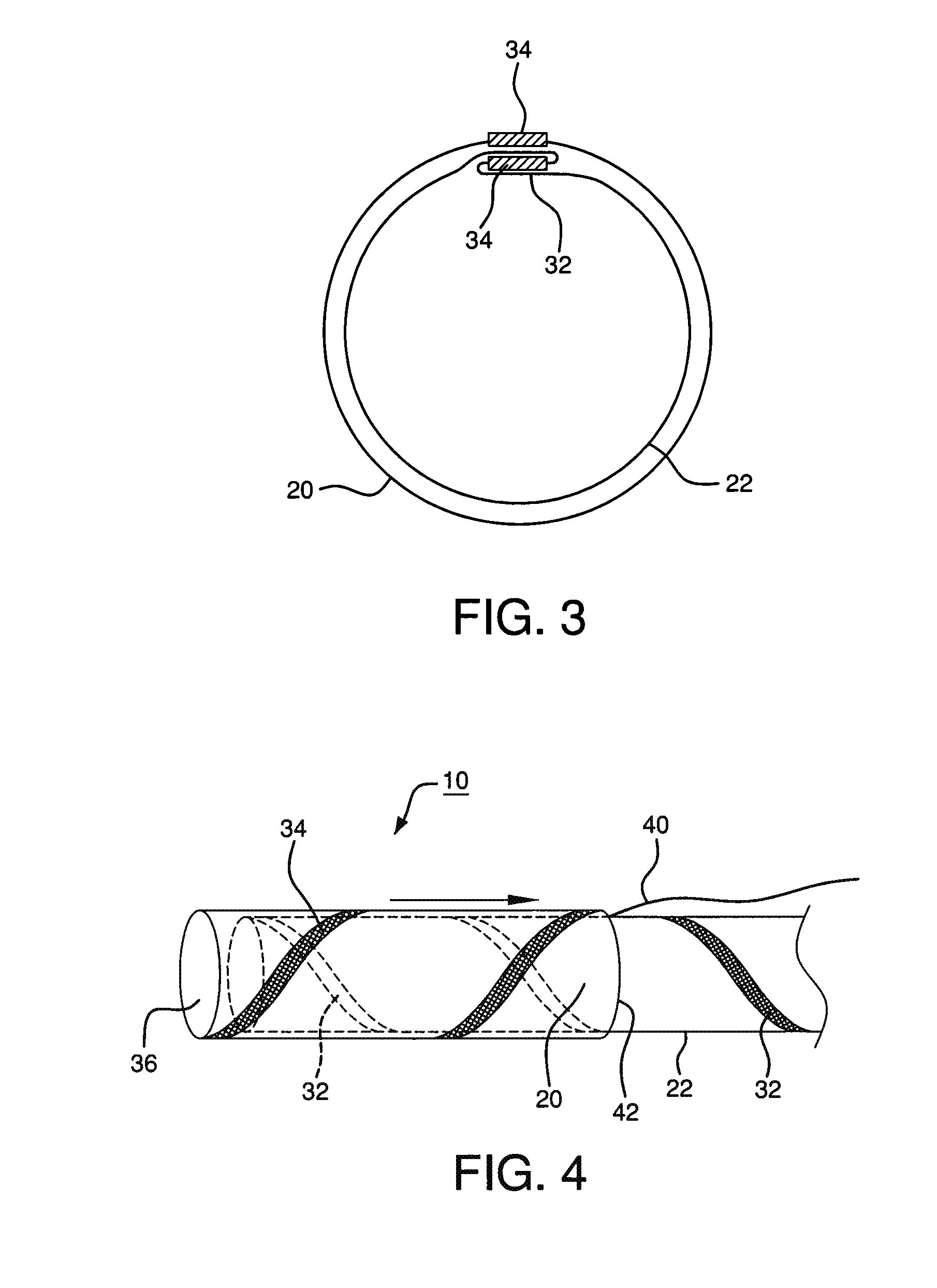
Pleated deployment sheath
a deployment sheath and plenum technology, applied in the field of plenum sheaths, can solve the problems of affecting the precise placement of the sheath, requiring otherwise unnecessary further treatment, and requiring risky conversion to open surgery, so as to facilitate the removal of the sheath, relieve friction of the everted sheath sliding along itself, and reduce the effect of tension
- Summary
- Abstract
- Description
- Claims
- Application Information
AI Technical Summary
Benefits of technology
Problems solved by technology
Method used
Image

Examples
example
[0080]Without intending to limit the scope of the present invention, the following example illustrates one embodiment of how the present invention may be practiced.
[0081]Sheath Tube Construct:
[0082](1) A 1″ (25.4 mm) wide strip of expanded polytetrafluoroethylene (ePTFE) film (having predominantly longitudinally oriented strength, film thickness of approximately 0.006 mm and break strength of approximately 0.8 kg / cm width) was “cigarette” wrapped on a 0.11″ (2.8 mm) diameter×40 cm long mandrel. The film structure was orientation parallel to mandrel axis so that the film was stronger parallel to the mandrel's longitudinal axis.
[0083](2) A second layer of 0.4″ (10 mm) wide ePTFE / FEP laminate film (predominantly longitudinally oriented strength, total film thickness of approximately 0.003 mm, FEP thickness of approximately 0.001 mm and break strength of approximately 0.7 kg / cm width) was then helically overwrapped around the first film layer with a single pass at a 0.2″ (5 mm) pitch, t...
PUM
 Abstract
Description
Claims
Application Information