Puncture resistant catheter for sensing vessels and for creating passages in tissue

a sensing vessel and catheter technology, applied in the field of catheters, can solve the problems of escalating complications, significant bleeding, and risks in many types of scope-based procedures, and achieve the effect of improving the ability of the sensing assembly and improving the ability to see the tip

Inactive Publication Date: 2009-08-13
BRONCUS MEDICAL
View PDF47 Cites 95 Cited by
  • Summary
  • Abstract
  • Description
  • Claims
  • Application Information

AI Technical Summary

Benefits of technology

[0011]In variations of the device, the sensing assembly is offset from an axis of the catheter assembly. The sensing assembly may also be positioned distal to the main catheter lumen. Doing so improves the ability of the sensing assembly to contact tissue surfaces when the device is advanced along body conduits. In addition, this offset and / or extension feature improves the ability to see the tip of the sensing assembly when the device is used with a scope type device.
[0012]The invention further includes methods of treating tissue, where the method includes selecting an area in the tissue for treatment, advancing the device into the lung to a tissue site, where the device includes a sensing assembly affixed to a catheter to sense for the presence or absence of blood vessels. The device may then allow for the use of a needle member comprising a curved needle that creates a passage. The device also includes various depth limiting features, such as a transition surface that causes tactile or sensory feedback to a physician if the device is advanced too far out the distal end of the sensing assembly, which can act as a stop. In addition, the needle assembly can include visual indicators to allow a physician to observe proper advancement of the needle.

Problems solved by technology

When performing procedures through an endoscope, bronchoscope, or other such device there is a risk that the procedure might disrupt structures beneath a tissue surface (such as blood vessels), where the disruption then causes significant complications.
One such area is within the airways of the lungs where puncturing of a blood vessel beneath the airway surface can result in significant bleeding.
In cases where a scope type device is used, the bleeding obstructs the ability of the medical practitioner to visualize the damaged area resulting in an escalation of complications.
Such risks occur in many types of scope-based procedures, including but not limited to lung based approaches.
For example, creation of collateral channels in COPD patients poses such risks.
In addition, biopsy procedures, and transbronchial aspiration procedures arc a few procedures that present the same risk of penetrating a blood vessel within the lungs.
The problem is further compounded when accounting for motion of the tissue.
For example, because airway or other lung tissue moves due to tidal motion of the lungs (as a result of the mechanics of breathing), it is difficult to visually identify an area that was previously scanned unless the scanning device remains relatively stationary against the tissue.
Moreover, the difficulty increases when considering that the procedure takes place through the camera of a bronchoscope or endoscope.
Aside from the risk to the patient, once the medical practitioner punctures a blood vessel, that practitioner is often understandably hesitant or risk averse when performing future procedures.
As a result, while the benefit of these procedures is well known, the risks of complications may reduce the overall success of the procedure.
In the case of a catheter carrying an extendable needle, and being urged through a working channel of a bronchoscope, a number of challenges may arise including, for example, increased friction between the needle and the catheter's working lumen.
The needle may also damage the bronchoscope, or worse, injure the patient.

Method used


Image

  • Puncture resistant catheter for sensing vessels and for creating passages in tissue
  • Puncture resistant catheter for sensing vessels and for creating passages in tissue
  • Puncture resistant catheter for sensing vessels and for creating passages in tissue

Examples


Embodiment Construction

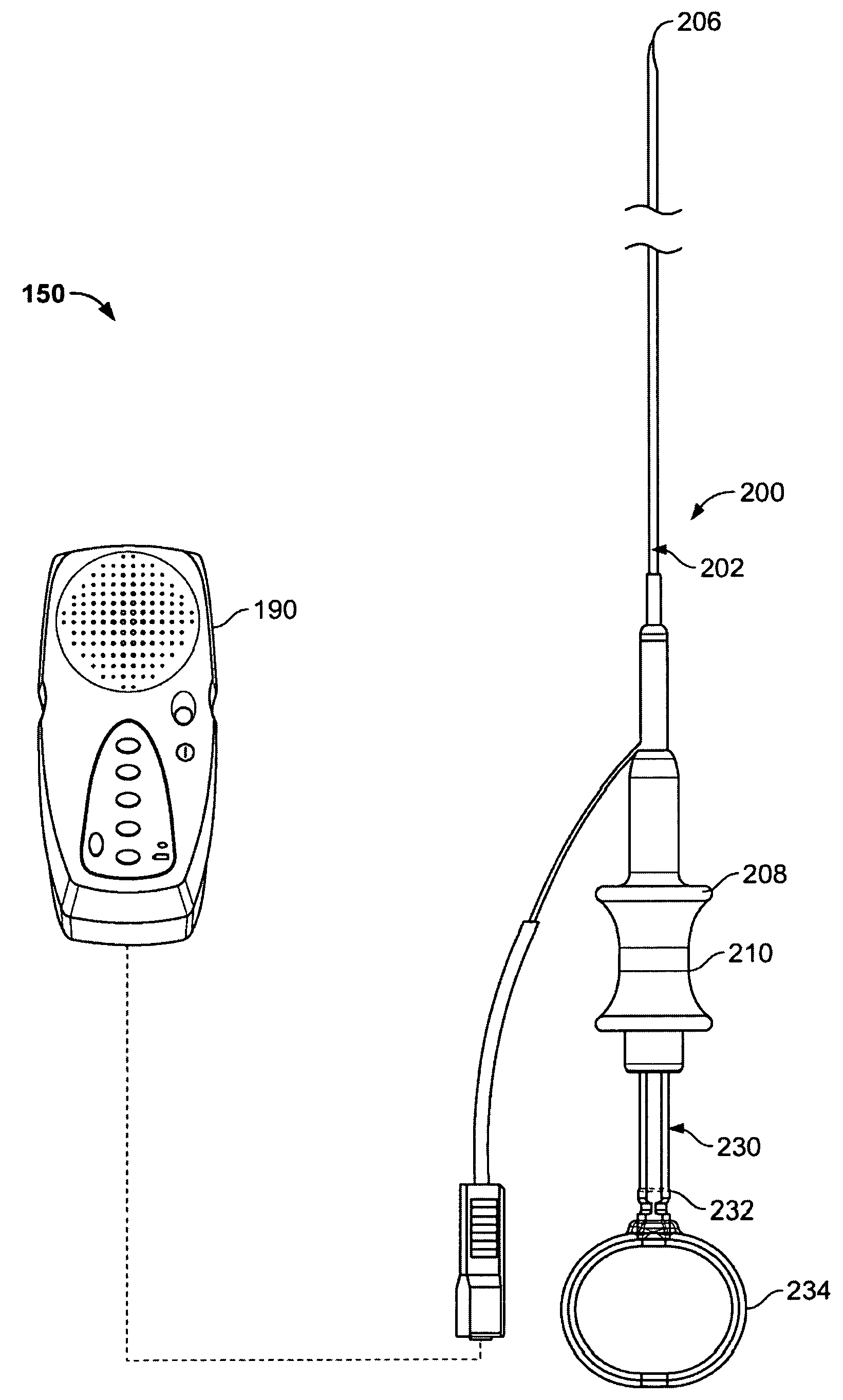
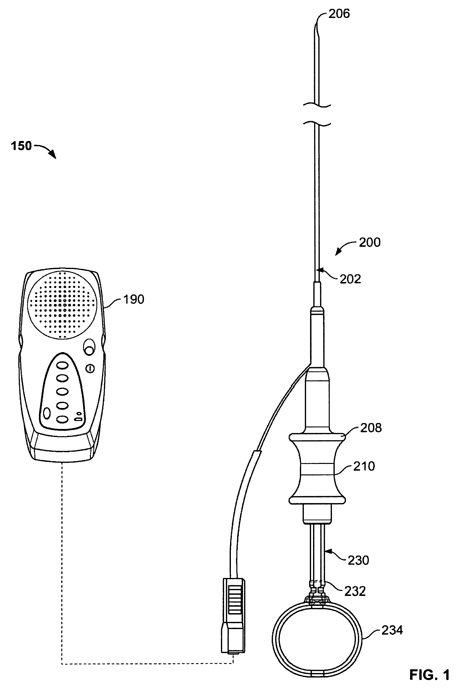
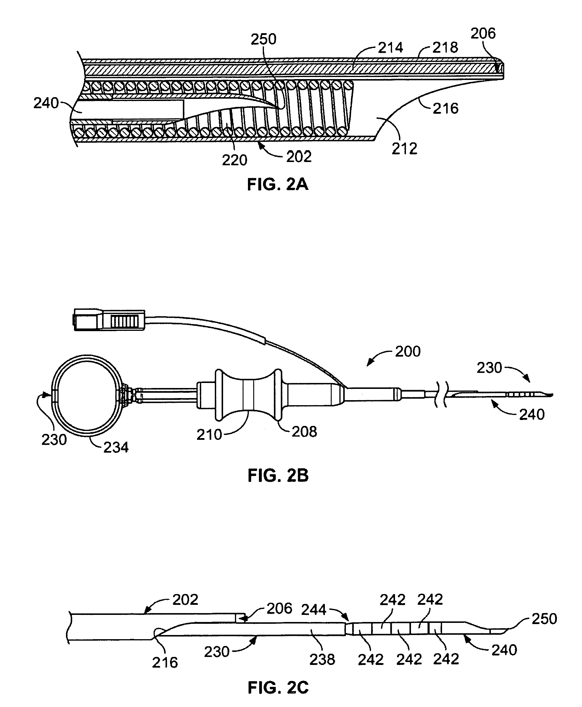
[0027]FIG. 1 illustrates a view of a variation of an inventive system 150 incorporating various features as described herein. The system generally includes a device 200 having an elongate sheath 202 with a sensing element 206 at a far end of the sheath 202. The far end of the sheath 202 shall be atraumatic so that movement of the sensing element 206 across tissue does not cause damage to the tissue. The elongate sheath 202 includes a working device extending therethrough. In this variation, the working device comprises a needle assembly 230. The device 200 is also coupled to (or connectable to) a control system 190 that is configured to assist the physician in scanning the tissue site. In the present variation, the control system 190 assists the physician in detecting whether blood vessels are at or near a particular target site. The control system 190 may be any type of unit that confirms the presence or absence of blood vessels. As such, it may be a thermal based system (incorpora...


PUM


Abstract

Devices and methods are disclosed for providing a sensing element to scan tissue and detect the presence of structures within the tissue to avoid the structures when performing a procedure on the tissue. A tissue penetrating member or needle is extendable from the device to create a channel or opening through the wall of an airway. The device is sufficiently flexible to navigate turns of at least 90 degrees. The distal section of the device includes puncture resistant sections, a curved needle tip, and depth limiting features that serve to create a channel in the lumen wall without causing collateral damage to the instruments, or patient.

Description

FIELD OF THE INVENTION[0001]The invention is directed to devices for sensing movement within tissue at a target site to scan for the presence or absence of structures such as blood vessels so that a procedure may be performed at the target site in a safe manner.BACKGROUND OF THE INVENTION[0002]When performing procedures through an endoscope, bronchoscope, or other such device there is a risk that the procedure might disrupt structures beneath a tissue surface (such as blood vessels), where the disruption then causes significant complications.[0003]One such area is within the airways of the lungs where puncturing of a blood vessel beneath the airway surface can result in significant bleeding. In cases where a scope type device is used, the bleeding obstructs the ability of the medical practitioner to visualize the damaged area resulting in an escalation of complications. In some cases, a patient's chest must be opened to stem the bleeding.[0004]Such risks occur in many types of scope...

Claims


Application Information

Patent Timeline
Patent Type & Authority Applications(United States)
IPC IPC(8): A61B8/14A61M25/06
CPCA61B1/018A61B1/2676A61B5/0059A61B5/489A61M2025/009A61M25/005A61M25/0068A61M25/0082A61B8/12
Inventor KEAST, THOMAS M.GWERDER, ERIC J.WEISS, AARON M.
Owner BRONCUS MEDICAL
Why Eureka
Try Eureka