Image acquisition for dual energy imaging

Active Publication Date: 2009-08-20
CARESTREAM HEALTH INC
View PDF9 Cites 7 Cited by
  • Summary
  • Abstract
  • Description
  • Claims
  • Application Information

AI Technical Summary

Benefits of technology

[0011]According to another aspect of the invention, there is provided a method acquiring dual energy x-ray images. The method includes providing a computer to control the acquisition of first and second x-ray images of a subject using an x-ray detector and an x-ray source to irradiate the subject. A filter is positioned between the x-ray source and the subject during the acquisition of the first and second images of the subject, the filter being selected of a filter material range Zf

Problems solved by technology

Lung cancer presents an burden to society because survival is low for advanced stage disease.
Conventional chest radiography has proven inadequate in the detection of early-stage disease, missing 50% of nodules measuring 10 mm or less.
Low-dose CT (LDCT) offers some improvement in di

Method used


Image

  • Image acquisition for dual energy imaging
  • Image acquisition for dual energy imaging
  • Image acquisition for dual energy imaging

Examples


Embodiment Construction

[0022]The following is a detailed description of the preferred embodiments of the invention, reference being made to the drawings in which the same reference numerals identify the same elements of structure in each of the several figures.

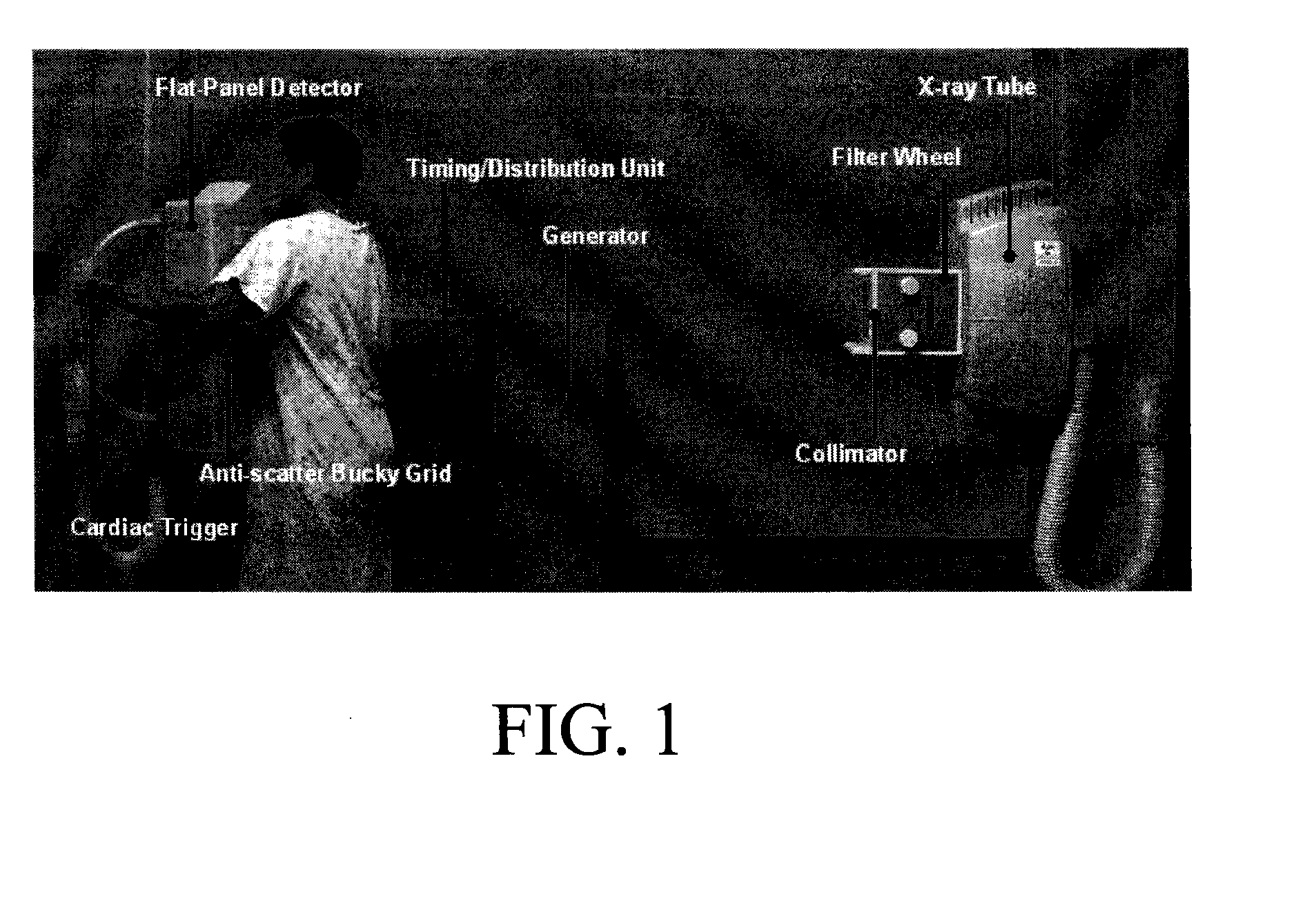
[0023]An exemplary dual energy (DE) imaging system is illustrated in FIG. 1. The system is based on a radiographic chest stand (RVG 5100 system, Eastman Kodak Company, Rochester, N.Y.), modified to perform cardiac-gated DE imaging.

[0024]The system includes a high-frequency, 3-phase generator (VZW 293ORD3-03, CPI, Georgetown, Ontario), a 400 kHU x-ray tube (Varian Rad-60, Salt Lake City, Utah), and a 10:1 antiscatter Bucky grid (Advanced Instrument Development Inc., Melrose Park, N.J.). Modifications to the RVG 5100 platform include:

[0025]1.) a collimator (Ralco R302 ACS / A, Biassono, Italy) incorporating a computer-controlled filter-wheel;

[0026]2.) a high-performance flat-panel detector, FPD (Trixell Pixium-4600, Moirans, France);

[0027]3.) a cardiac ...


PUM


Abstract

Acquisition techniques for dual energy (DE) chest imaging system. Technique factors include the added x-ray filtration, kVp pair, and the allocation of dose between low- and high-energy projections, with total dose equal to or less than that of a conventional chest radiograph. Factors are described which maximize lung nodule detectability as characterized by the signal difference to noise ratio (SDNR) in DE chest images. kVp pair and dose allocation are described using a chest phantom presenting simulated lung nodules and ribs for thin, average, and thick body habitus. Low- and high-energy techniques ranged from 60-90 kVp and 120-150 kVp, respectively, with peak soft-tissue SDNR achieved at [60/120] kVp for patient thicknesses and levels of imaging dose. A strong dependence on the kVp of the low-energy projection was observed.

Description

CROSS REFERENCE TO RELATED APPLICATIONS[0001]Reference is made to, and priority is claimed from, commonly assigned application U.S. Ser. No. 61 / 028,950, entitled OPTIMIZAION OF IMAGE ACQUISITION TECHNIQUE FOR DUAL-ENERGY IMAGING OF THE CHEST, and filed on Feb. 15, 2007 in the name of Van Metter, incorporated herein by reference.FIELD OF THE INVENTION[0002]The invention relates generally to the field of dual energy projection radiography, and in particular to dual energy imaging of the chest.BACKGROUND OF THE INVENTION[0003]In multiple-energy projection radiographic imaging, a number of images of the same object are acquired that reveal the x-ray transmittance of the object for differing x-ray spectra. In dual energy imaging, two images of the same object are acquired sequentially under different x-ray beam conditions, such as beam energy and filtration. These images are proportional to the x-ray transmittance of the object for the differing x-ray spectra. These images can then be de...

Claims


Application Information

Patent Timeline
IPC IPC(8): H05G1/60
CPCA61B6/4035A61B6/482A61B6/541A61B6/503A61B6/505A61B6/50
Inventor SHKUMAT, NICK A.SIEWERDSEN, JEFFREY H.YORKSTON, JOHNVAN METTER, RICHARD L.
Owner CARESTREAM HEALTH INC
Why Eureka
Try Eureka