[0009]In an embodiment, this need is also addressed by a computer-implemented method of analyzing one or more mammograms. The method includes determining a contour line, which divides an object area defined by an object from a background area of a mammogram, and automatically positioning and scaling the mammogram by means of the contour line, to position of the mammogram relative to another mammogram and to reduce the mammogram's background area relative to the object area.
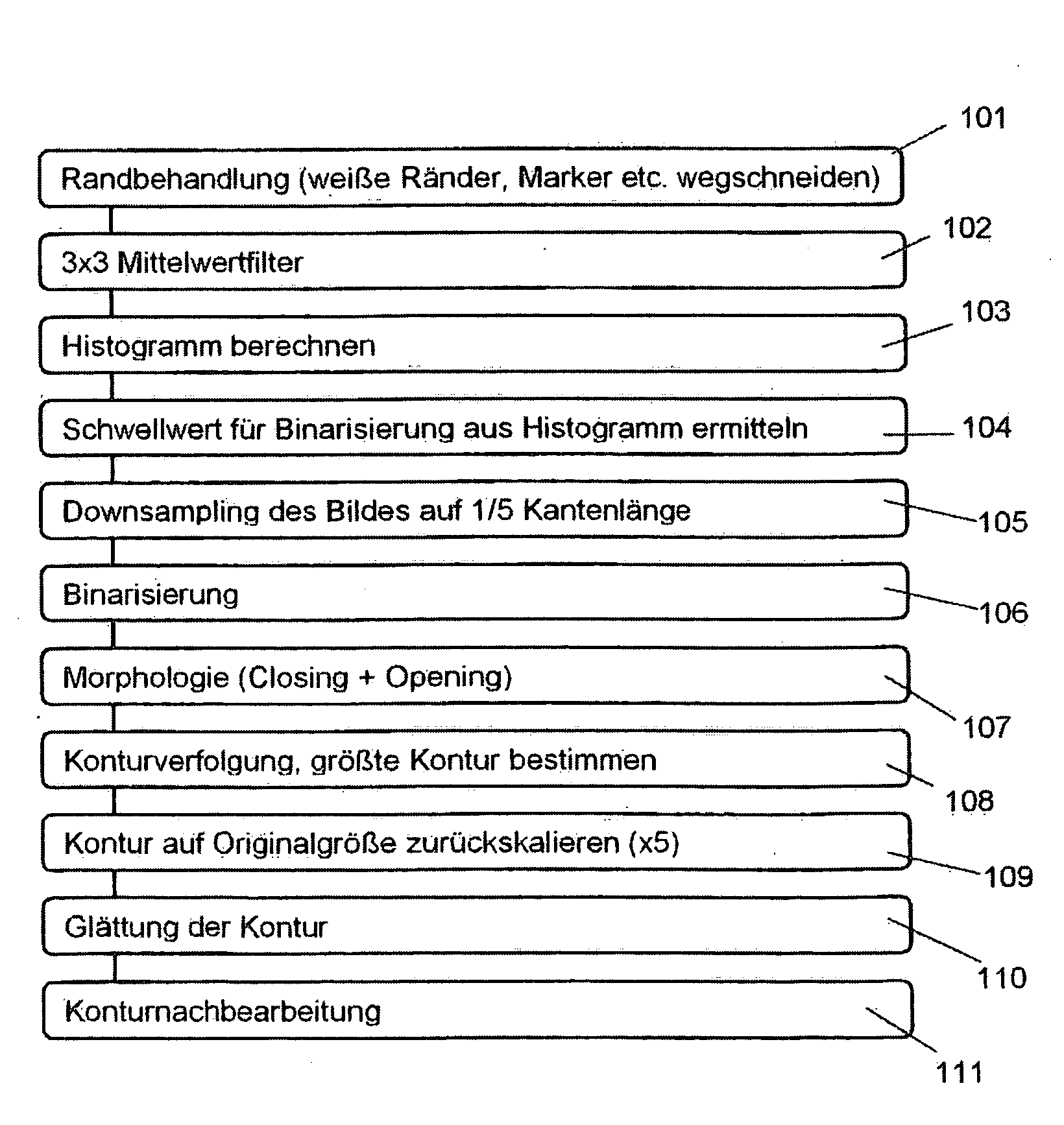
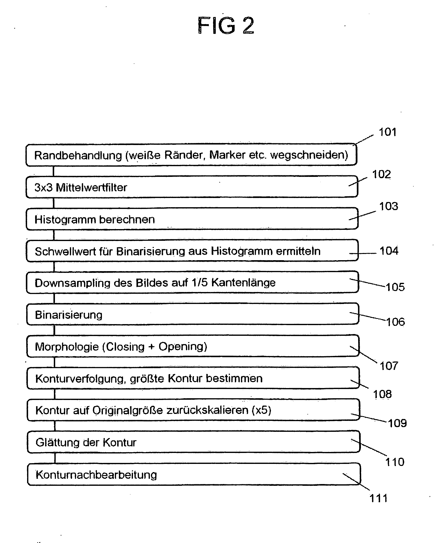
[0014]For the detection of the contour line, a method can be used advantageously, so that depending on the image characteristic, thus, for example, depending on the bit-depth, photo type, the manufacturer and the
image noise, of the respective mammogram to be processed, a threshold method, with which at least one threshold is used for the binarization of the mammogram, and / or a
scan line method, with which the gradient of the mammogram is determined along different scan lines, is used for the detection of a contour line of the mammogram surrounding an object area. The knowledge which underlies this is, that with mammograms differing strongly in respect to the image characteristic, a uniform method for the detection of an object area is not sufficient, but rather, depending on the image characteristic, different methods have to be used, in order to reliably detect a contour line surrounding an object area of a mammogram. Therefore, a means is provided, by means of which different methods can be applied alternatively or in combination to a mammogram, in order to determine the contour line surrounding the object area of the mammogram in a reliable and robust manner. The proposed method for the detection of the contour line offers an
advantage, that mammograms differing in their image characteristic, which can vary particularly with regard to the bit-depth, the
dynamic range, gray-scale value distribution and the
noise, for example, depending on the manufacturer of the X-
ray machine used and the photo-modality, can be processed, in which—independent of the type of mammogram—a uniform output occurs in the form of a contour line delineating the object area or of a masking image generated by means of a contour line.
[0028]Both with the different variants of the threshold method or with the
scan line method it can be advantageously provided, to filter the mammogram by means of an average value filter operating locally on a group of pixels of the mammogram before the actual detection of the contour line. Such average value filters serve to form an average value locally in an area of the mammogram, for example, for a group of 3×3 or 7×7 pixels and thus to smear over and smooth the gray-scale value distribution of the mammogram, so that high-
frequency noise components are suppressed and removed from the mammogram.
[0029]In a further design of the method, based on the determined contour line of the object displayed by the mammogram in the case of an interactive input of a finding by a doctor into the mammogram, the coordinates of the input can be determined and be transformed into a desired output format. Basically, in this manner it should be made possible for a doctor to input a finding directly in the indicated mammogram, through the analysis of the mammogram to automatically process, display and save the information contained in the finding. In particular, it is in this connection advantageous, if the coordinates of the input of the doctor are transformed into a time-of-day model, by means of which the position of the finding in the mamma can be indicated and displayed in an explicit and clearly laid out manner.

