
Imaging support device for radiographic long length imaging

a support device and imaging technology, applied in the field of imaging support devices for radiographic long length imaging, can solve the problems of inconvenient image combination, achieve the effect of reducing the length of images, enhancing work efficiency in long length imaging, and reducing the failure rate of image combination alignmen

Inactive Publication Date: 2009-09-24
FUJIFILM CORP
View PDF5 Cites 24 Cited by
  • Summary
  • Abstract
  • Description
  • Claims
  • Application Information

AI Technical Summary

Benefits of technology

The present invention provides an image taking support device for radiographic long length imaging that can easily arrange markers and reduce the time for image taking. The device includes a supporting member, markers, an irradiated area determination device, a marker position determining device, and a marker moving device. The markers can be arranged parallel to the subject or on two rod-like members that can be moved in parallel to each other. The device can automatically determine the area to be irradiated and position the markers accordingly, reducing the failure of alignment for image combination and improving the quality of long length images. The device also eliminates the need for an image-taking person to set the markers, saving time and improving work efficiency.

Problems solved by technology

Where performing long length imaging involving taking plural images as described above, the posture of a subject (patient) may change during the X-ray detector and the X-ray tube moving between image takings, causing inconvenience for image combination.

Method used


Image

  • Imaging support device for radiographic long length imaging
  • Imaging support device for radiographic long length imaging
  • Imaging support device for radiographic long length imaging

Examples


first embodiment

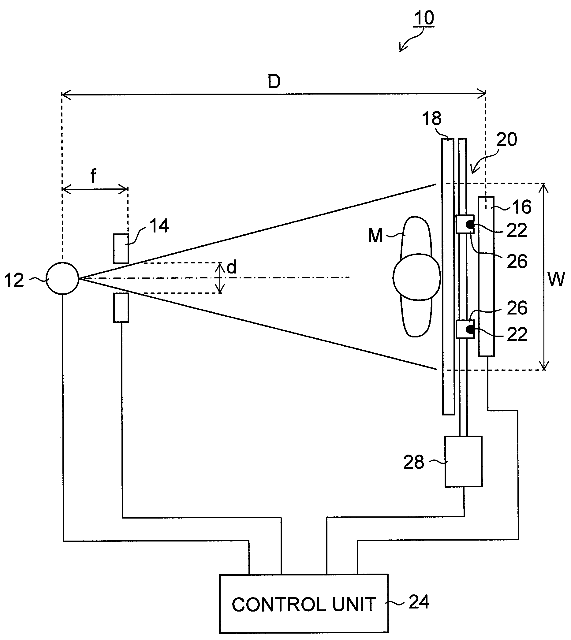
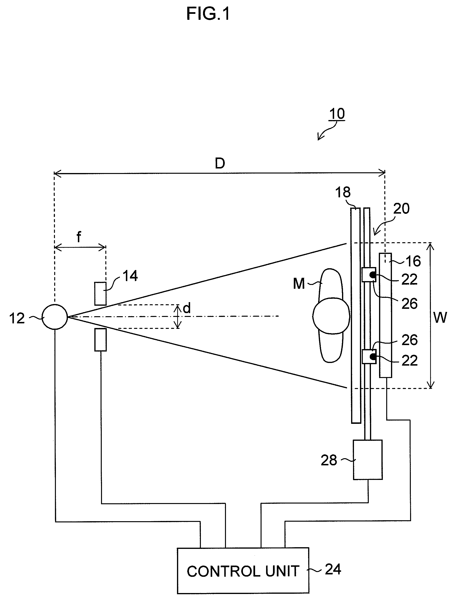
[0035]FIG. 1 is a schematic diagram of a configuration of a radiographic apparatus using an image taking support device for radiographic long length imaging according to the present invention.

[0036]As illustrated in FIG. 1, a radiographic apparatus 10 according to the present embodiment mainly includes: an X-ray source 12 that applies an X-ray to a subject M; a collimator 14 that focuses an X-ray emitted from the X-ray source 12; a flat panel-type X-ray detector (FPD: flat panel detector) 16 that detects an X-ray that has passed through the subject M and outputs detection signals; a marker device 20 including a screen 18 and markers 22 that provide marks for combining plural images, which is included in a taking support device for long length imaging; and a control unit 24 that controls the aforementioned components.

[0037]Although a description of the detailed configuration will not be provided, the X-ray source 12 includes an X-ray tube that applies an X-ray to the subject M, and a...

second embodiment

[0075]Next, the present invention will be described.

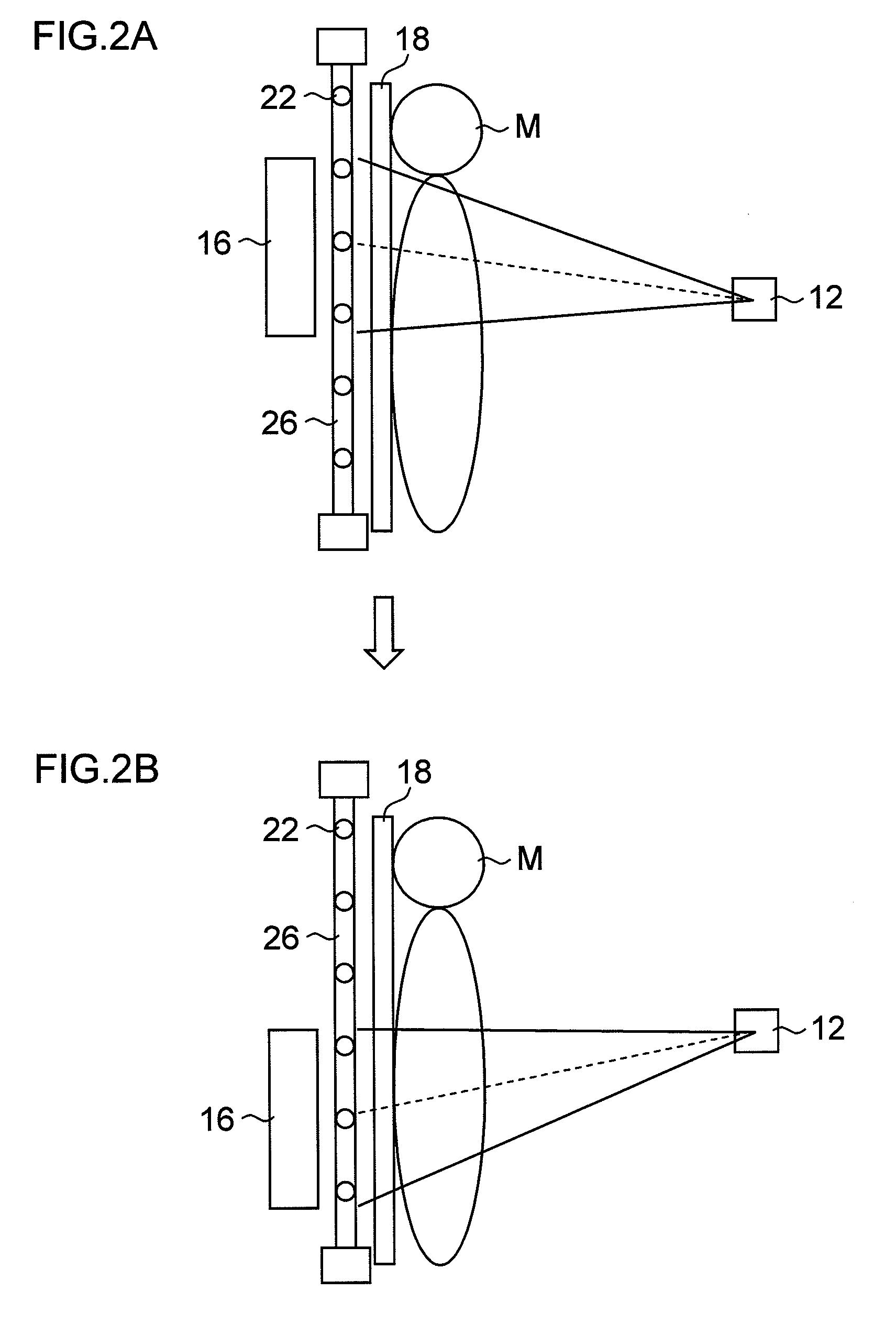
[0076]In the present embodiment, an irradiation field lamp that emits visible light is provided at a position that is substantially the same as the position of the X-ray source, each marker holding device is provided with a photosensor to automatically determine a field of view width W, and markers are automatically moved.

[0077]FIG. 6 illustrates a schematic diagram of a configuration of a radiographic apparatus using an image taking support device for radiographic long length imaging according to the present embodiment.

[0078]As illustrated in FIG. 6, the image taking support device according to the present embodiment includes an irradiation field lamp 150 provided at a position that is substantially the same as the position of an X-ray source 112, the irradiation field lamp 150 emitting visible light; photosensors 152 provided on marker holding members 126 separately from markers 122, the photosensors 152 being arranged at regular...


PUM


Abstract

An image taking support device for radiographic long length imaging, the image taking support device providing an image of a subject, the image being longer than a length of a maximum field of view of a radiation detector, by combining a plurality of radiographic images each partially overlapping another at the overlapping parts, the device comprising:a supporting member including a radiotransparent material and supporting the subject;markers including a substance with a large radiation attenuation coefficient, the markers being movably arranged along a surface of the supporting member supporting the subject so that the markers provide marks for combining the images;an irradiated area determination device that determines an area to be irradiated with a radiation;a marker position determining device that determines positions for arranging the markers, based on the determined area to be irradiated; anda marker moving device that moves the markers to the determined positions of the markers.

Description

BACKGROUND OF THE INVENTION[0001]1. Field of the Invention[0002]The present invention relates to an image taking support device for radiographic long length imaging, and specifically relates to an image taking support device for radiographic long length imaging using an FPD (flat panel detector), the device being used for, when connecting a plurality of images obtained by performing image-taking while changing the radiation field of view, correctly aligning the connection positions of the images.[0003]2. Description of the Related Art[0004]Conventionally, radiographic apparatuses that apply radiation, such as an X-ray, to a subject and detect radiation passing through the subject to obtain a radiographic image for diagnosis have widely been known. Among these radiographic apparatuses, for example, one using a radiographic film, or a storage phosphor sheet that stores radiation energy, as a radiation detector that detects radiation passing through the subject has been known.[0005]Als...

Claims


Application Information

Patent Timeline
Patent Type & Authority Applications(United States)
IPC IPC(8): H05G1/28A61B6/08
CPCA61B6/04A61B6/5241A61B6/4283A61B6/4233
Inventor KAWAMURA, TAKAHIROYAMAGUCHI, YOSHITAKA
Owner FUJIFILM CORP
Learn More