Treatment instrument for endoscope

a treatment instrument and endoscope technology, applied in the direction of surgical staples, catheters, osteosynthesis devices, etc., can solve the problems of long treatment time and huge technical difficulties

Inactive Publication Date: 2009-10-08
JICHI MEDICAL UNIVERSITY
View PDF8 Cites 7 Cited by
  • Summary
  • Abstract
  • Description
  • Claims
  • Application Information

AI Technical Summary

Benefits of technology

[0029]According to the present invention, since a treatment section has a side surface that has a sandwich structure in which a strip-shaped conductor is sandwiched between non-conductors, a cutting of subm

Problems solved by technology

The method enables a drawing of an incision line as intended, and ensures the extirpation of tumor, but at the same time, it involves tremendous technical

Method used


Image

  • Treatment instrument for endoscope
  • Treatment instrument for endoscope
  • Treatment instrument for endoscope

Examples


first embodiment

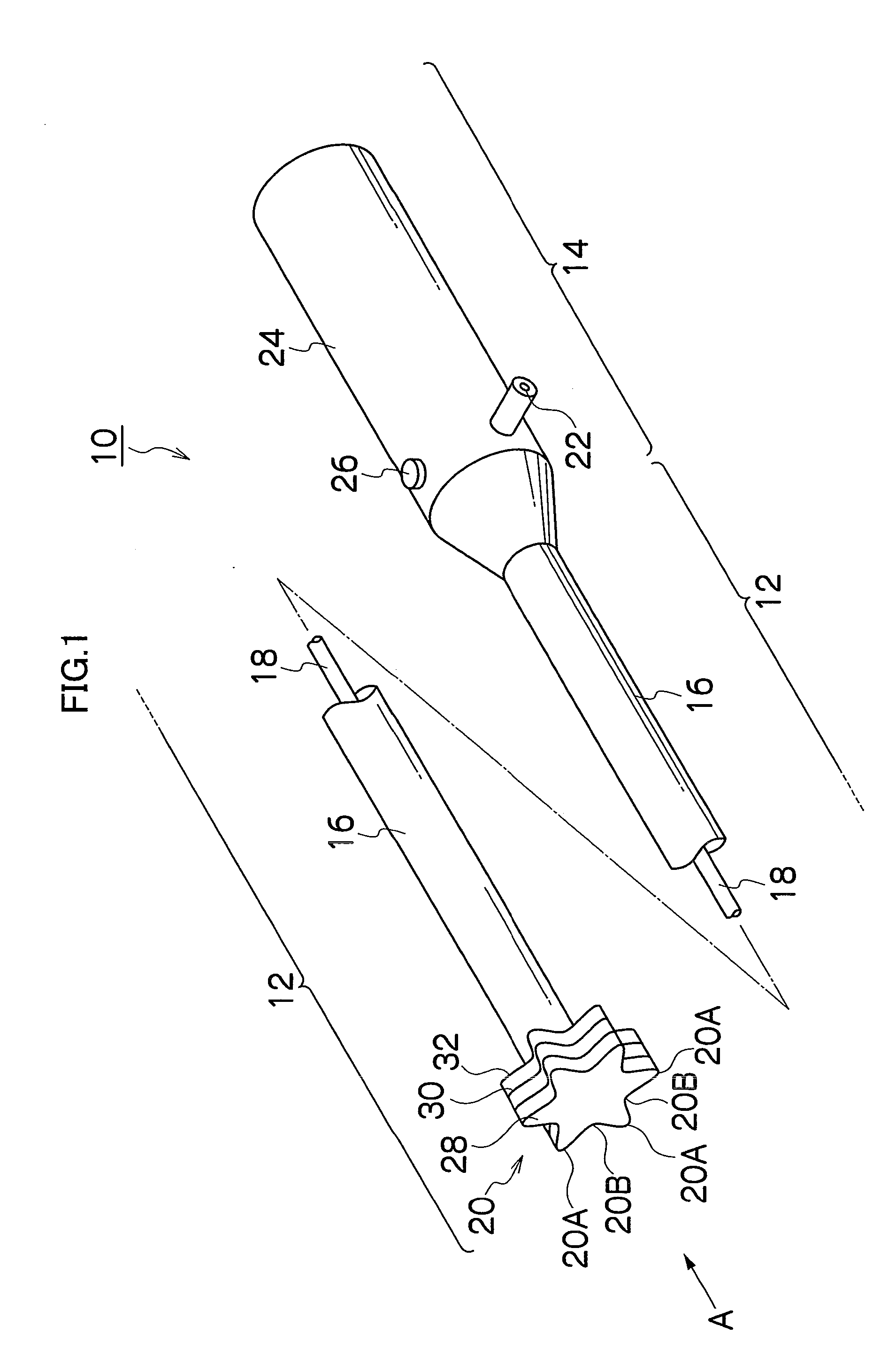
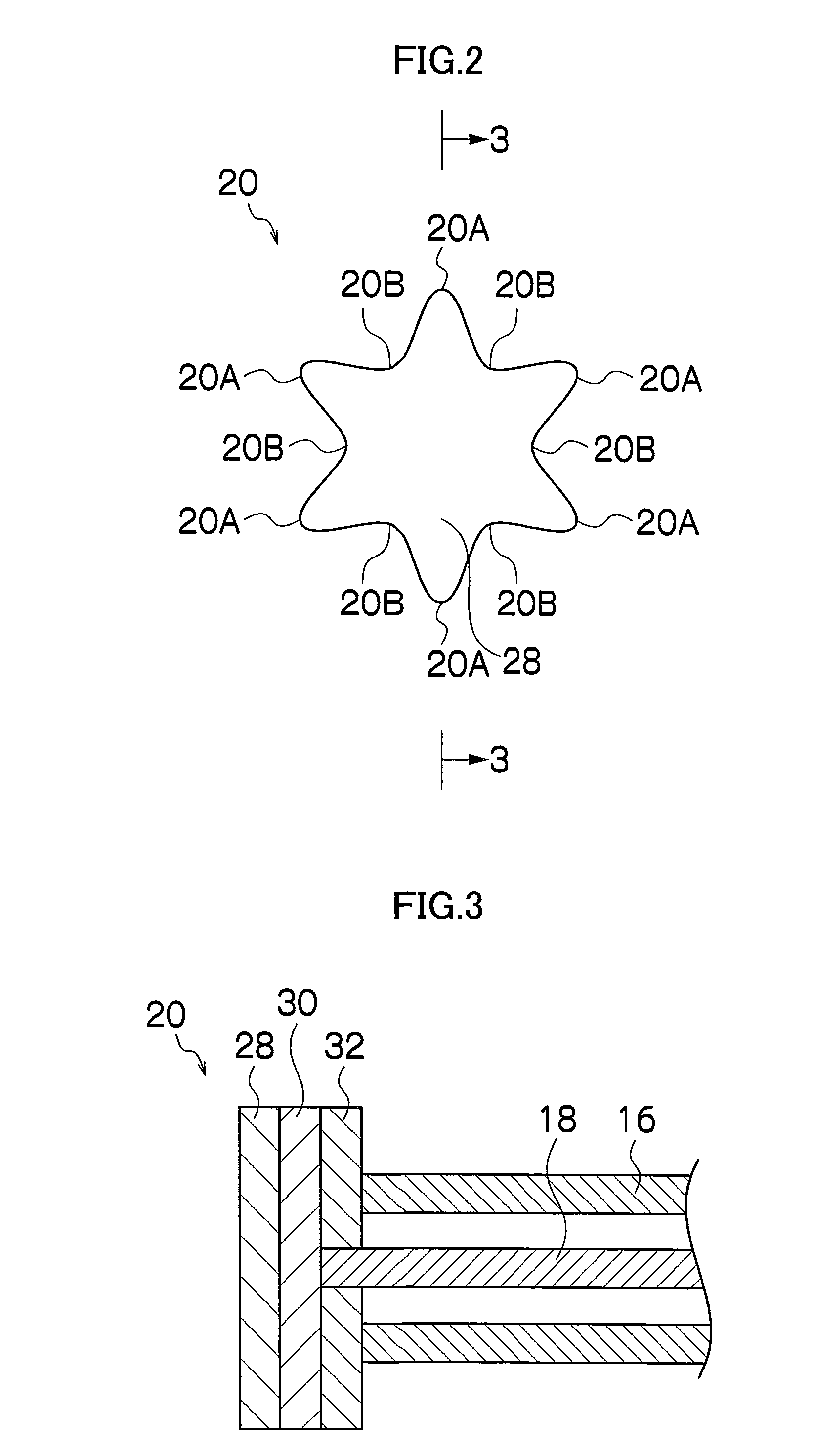
[0061]FIG. 1 is a perspective view showing a treatment instrument for endoscope 10. The treatment instrument for endoscope 10 generally includes, as shown in FIG. 1, an insertion section 12 which is inserted into a body, and a hand-side operation section 14 connected to the insertion section 12, and the insertion section 12 is configured with a non-conductive and flexible sheath 16, a conductive wire 18 which is inserted into the sheath 16, and a treatment section 20 mounted to a distal end of the flexible sheath 16. The distal end of the wire 18 is connected to the treatment section 20, and the proximal end of the wire 18 is connected to a connector 22 of the hand-side operation section 14. The connector 22 is electrically connected to a high-frequency current supply apparatus (not shown) for supplying a high frequency current. The hand-side operation section 14 has a grasping portion 24 with an operation button 26, so that a press-down of the operation button 26 causes a high freq...

second embodiment

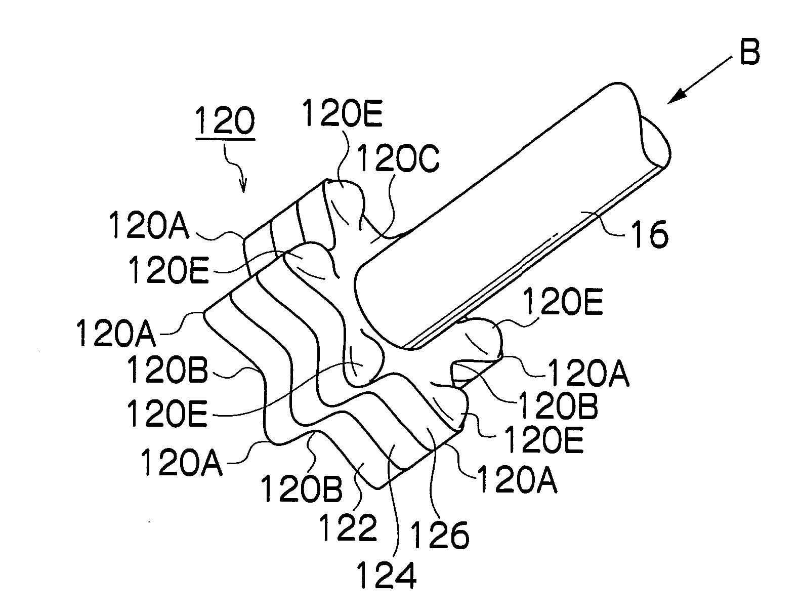
[0084]FIG. 7 is a side view showing the distal end portion of a treatment instrument for endoscope of a second embodiment, FIG. 8 is a side cross sectional view showing the cross section of FIG. 7, FIG. 9 is a side view showing the treatment section of FIG. 7 in another posture, and FIG. 10 is a view of the treatment section of FIG. 9 seen in the direction B.

[0085]As shown in these figures, in the second embodiment, the treatment section 20 has a proximal end surface 20C from which a pair of bearings 42, 42 are protruded, and the bearings 42, 42 support a shaft 44. To the shaft 44, a first sleeve 46 is rotatably coupled at the distal end portion thereof, and to the proximal end portion of the first sleeve 46, a second sleeve 48 is engaged at the distal end portion thereof. The first sleeve 46 and the second sleeve 48 are coaxially arranged and coupled with each other, and also are coupled to relatively rotate about the central axes of the sleeves. To the proximal end of the second s...

third embodiment

[0101]The third embodiment is a bipolar-type treatment instrument performing a coagulation process only on the tissues between the electrodes (between the conductors), which enables a control to narrow the tissue area for coagulation. Therefore, the tissue area which may be damaged is narrowed, and the tissue damage caused by coagulation can be minimized.

[0102]In the above described third embodiment, both of the distal end side (the proper muscular layer 36 side) and proximal end side (the mucous membrane 34 side) of a cutting is processed for coagulation, but the positions for coagulation are not limited to those, and for example, a high frequency current for coagulation may be applied to the part between the conductors 91 and 94 so as to coagulate the entire area. Alternatively, a high frequency current for coagulation may be applied only to the conductors 91 and 92, so as to coagulate only the distal end side of the incision position.

[0103]Also, in the above described third embod...


PUM


Abstract

A treatment instrument for endoscope that is suitable for cutting submucosal layer in endoscopic submucosal dissection is provided, and the treatment instrument for endoscope has a treatment section at a distal end of an insertion section which is inserted in a body. The treatment section includes a conductor that is connected to a high-frequency source, and is configured to have a sandwich structure in which the conductor is sandwiched between a non-conductor on the distal end side thereof and a non-conductor on a proximal end side thereof. The conductor is formed to be exposed in a strip-shape at a side surface of the treatment section, and a lateral movement of the treatment section causes a cutting of a submucosal layer with the conductor to be performed.

Description

TECHNICAL FIELD[0001]The present invention relates to a treatment instrument for endoscope, more particularly, to a treatment instrument for endoscope which is used in endoscopic submucosal dissection (ESD).BACKGROUND ART[0002]Endoscopic mucosal resection is known for its usefulness as a minimally invasive radical surgery that is a reliable treatment for neoplastic mucosal lesion such as early gastric cancer and early colorectal cancer. In recent years, a method called endoscopic submucosal dissection (ESD) has been developed and widely used as a reliable endoscopic demucosation method for en bloc resection of a wider lesion. The method includes an incision of mucous membrane around a tumor, and a cutting of a submucosal layer between the mucous membrane and a proper muscular layer for en bloc resection of tumor mucous membrane. The method enables a drawing of an incision line as intended, and ensures the extirpation of tumor, but at the same time, it involves tremendous technical d...

Claims


Application Information

Patent Timeline
IPC IPC(8): A61B18/18A61B17/94
CPCA61B1/018A61B18/1492A61B18/1815A61B2018/1861A61B2018/00482A61B2018/1405A61B2018/1497A61B2018/00083A61B17/00A61B17/32
Inventor YAMAMOTO, HIRONORI
Owner JICHI MEDICAL UNIVERSITY
Why Eureka
Try Eureka