Off-axis visualization systems

a visualization system and axis technology, applied in the field of medical devices, can solve the problems of affecting the fine positioning of the imaging system, cramping the working area created by such inflatable balloons, and reducing the available space within the catheter, so as to reduce or eliminate friction during deployment and retraction.

Active Publication Date: 2009-12-03
INTUITIVE SURGICAL OPERATIONS INC
View PDF107 Cites 81 Cited by
  • Summary
  • Abstract
  • Description
  • Claims
  • Application Information

AI Technical Summary

Benefits of technology

[0017]The imaging hood may be deployed into an expanded shape and retracted within a catheter utilizing various mechanisms. Moreover, the imaging element, such as a CCD or CMOS imaging camera, may be positioned distally or proximally of the imaging hood when collapsed into its low-profile configuration. Such a configuration may reduce or eliminate friction during deployment and retraction as well as increase the available space within the catheter not only for the imaging unit but also for the hood.
[0018]Moreover, the imaging element may be introduced along or within the hood into an off-axis position relative to a longitudinal axis of the catheter and / or hood for providing direct visualization of the underlying tissue to be visually examined and / or treated. For example, one variation may utilize a flexible section located at a distal end of the catheter which may be configured from various flexible materials coupled or integrated with a relatively rigid section located proximally of flexible section. The imaging element may be positioned and / or attached to a lateral inner wall of the flexible section such that when the section is collapsed within the sheath, the imaging element may be placed in an in-line or axially positioned relative to the catheter and hood to provide for a low-profile delivery configuration.
[0020]Because the imager is positioned laterally, the catheter and hood may accommodate a variety of sizes for different types of imagers. For instance, relatively larger, more economical, and / or relatively more powerful CCD or CMOS imagers may be utilized with the system as the hood may accommodate a range of sizes and configurations for the imaging system. With the imager positioned in its off-axis location relative to the hood and / or catheter, the user may obtain a better angle of visualization of the entire operating landscape, including both the movements of the tools and the target tissue surface during any number of therapeutic and / or diagnostic procedures. Moreover, the unobstructed opening of the catheter may allow for various instruments, such as RF ablation probes, graspers, needles, etc., to be deployed through the catheter and past the imager into the open area defined by the hood for treatment upon the underlying imaged tissue.

Problems solved by technology

However, such imaging balloons have many inherent disadvantages.
For instance, such balloons generally require that the balloon be inflated to a relatively large size which may undesirably displace surrounding tissue and interfere with fine positioning of the imaging system against the tissue.
Moreover, the working area created by such inflatable balloons are generally cramped and limited in size.
Furthermore, inflated balloons may be susceptible to pressure changes in the surrounding fluid.
For example, if the environment surrounding the inflated balloon undergoes pressure changes, e.g., during systolic and diastolic pressure cycles in a beating heart, the constant pressure change may affect the inflated balloon volume and its positioning to produce unsteady or undesirable conditions for optimal tissue imaging.
Accordingly, these types of imaging modalities are generally unable to provide desirable images useful for sufficient diagnosis and therapy of the endoluminal structure, due in part to factors such as dynamic forces generated by the natural movement of the heart.
Moreover, anatomic structures within the body can occlude or obstruct the image acquisition process.
Also, the presence and movement of opaque bodily fluids such as blood generally make in vivo imaging of tissue regions within the heart difficult.
However, such imaging modalities fail to provide real-time imaging for intra-operative therapeutic procedures.
However, fluoroscopy fails to provide an accurate image of the tissue quality or surface and also fails to provide for instrumentation for performing tissue manipulation or other therapeutic procedures upon the visualized tissue regions.
Moreover, many of the conventional imaging systems lack the capability to provide therapeutic treatments or are difficult to manipulate in providing effective therapies.
For instance, the treatment in a patient's heart for atrial fibrillation is generally made difficult by a number of factors, such as visualization of the target tissue, access to the target tissue, and instrument articulation and management, amongst others.
Conventional catheter techniques and devices, for example such as those described in U.S. Pat. Nos. 5,895,417; 5,941,845; and 6,129,724, used on the epicardial surface of the heart may be difficult in assuring a transmural lesion or complete blockage of electrical signals.
In addition, current devices may have difficulty dealing with varying thickness of tissue through which a transmural lesion desired.
Conventional accompanying imaging devices, such as fluoroscopy, are unable to detect perpendicular electrode orientation, catheter movement during the cardiac cycle, and image catheter position throughout lesion formation.
Without real-time visualization, it is difficult to reposition devices to another area that requires transmural lesion ablation.
The absence of real-time visualization also poses the risk of incorrect placement and ablation of critical structures such as sinus node tissue which can lead to fatal consequences.

Method used


Image

  • Off-axis visualization systems
  • Off-axis visualization systems
  • Off-axis visualization systems

Examples


Embodiment Construction

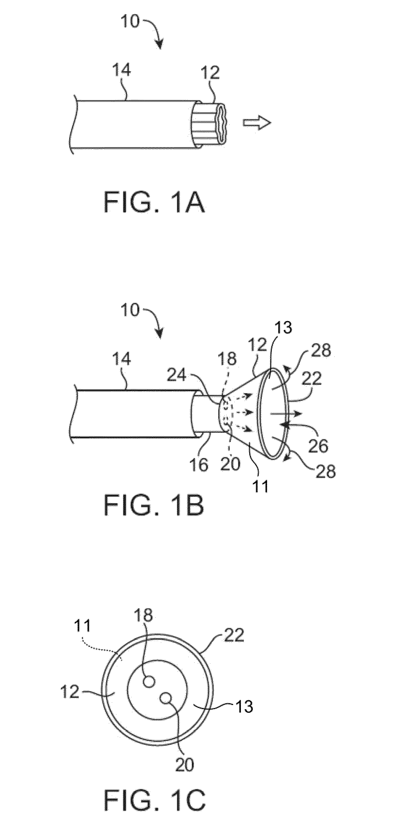
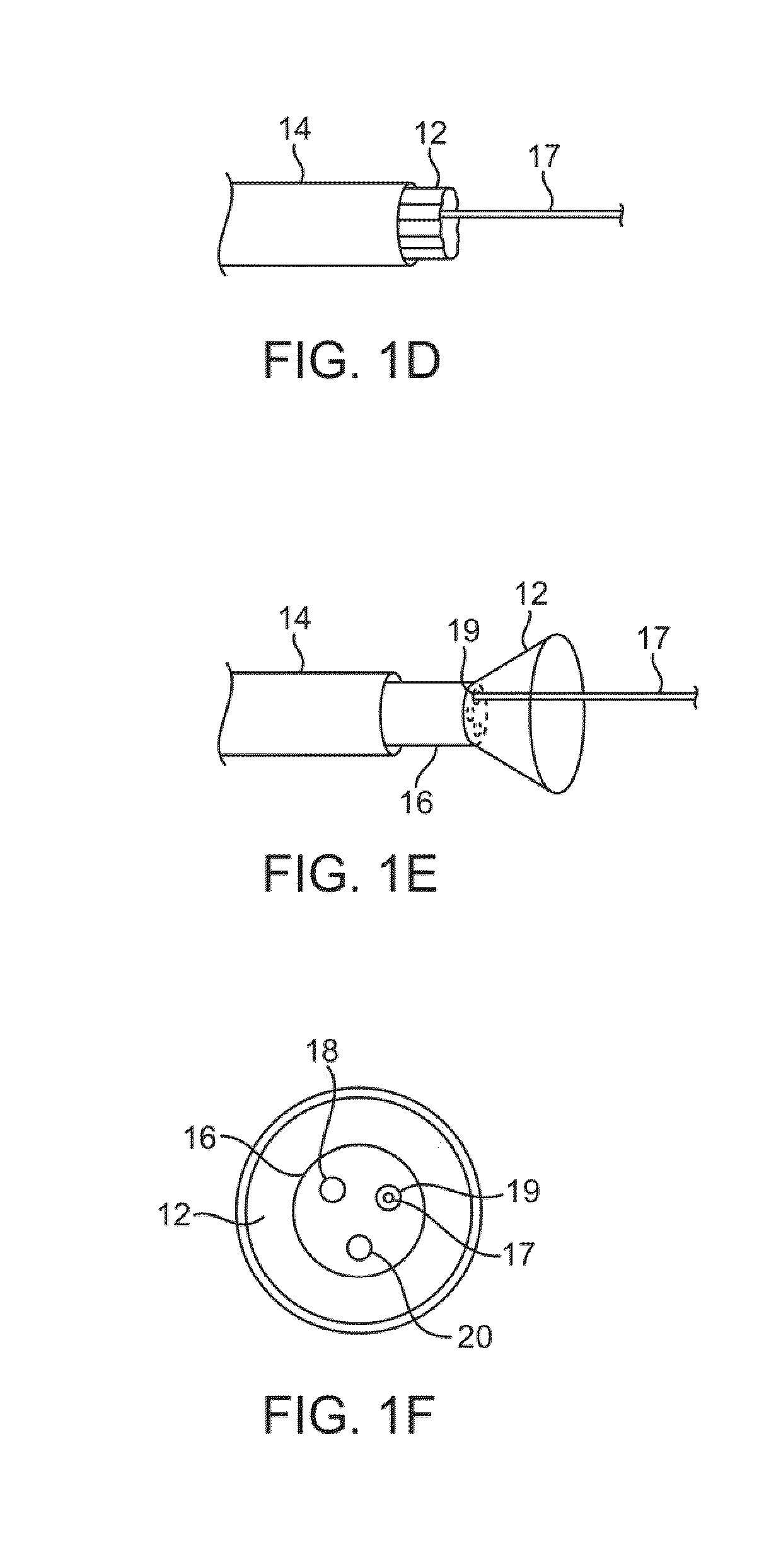
[0068]A tissue-imaging and manipulation apparatus described below is able to provide real-time images in vivo of tissue regions within a body lumen such as a heart, which is filled with blood flowing dynamically therethrough and is also able to provide intravascular tools and instruments for performing various procedures upon the imaged tissue regions. Such an apparatus may be utilized for many procedures, e.g., facilitating transseptal access to the left atrium, cannulating the coronary sinus, diagnosis of valve regurgitation / stenosis, valvuloplasty, atrial appendage closure, arrhythmogenic focus ablation, among other procedures. Further examples of tissue visualization catheters which may be utilized are shown and described in further detail in U.S. patent application Ser. No. 11 / 259,498 filed Oct. 25, 2005, which has been incorporated hereinabove by reference in its entirety.

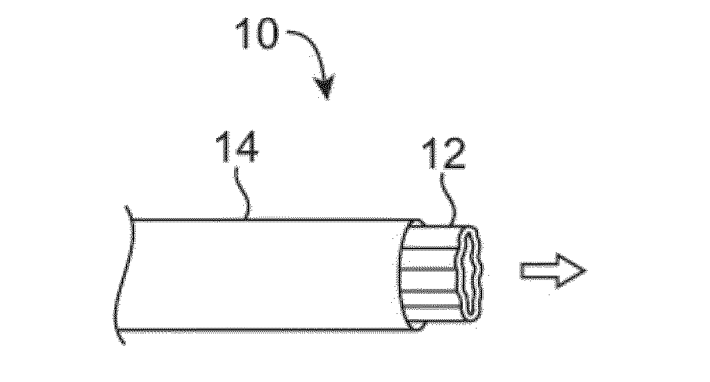
[0069]One variation of a tissue access and imaging apparatus is shown in the detail perspective views of F...


PUM


Abstract

Off-axis visualization systems are described herein which facilitate the deployment, visualization, and retraction of an imaging element from a catheter. Such a system may include a deployment catheter and an attached imaging hood deployable into an expanded configuration as well as an imaging element, such as a CCD or CMOS imager, which may be deployed from a low profile configuration into a position which is off-axis relative to a longitudinal axis of the deployment catheter and / or imaging hood.

Description

CROSS-REFERENCE TO RELATED APPLICATIONS[0001]This application claims the benefit of priority to U.S. Prov. Pat. Apps. 60 / 871,415 and 60 / 871,424 both filed Dec. 21, 2006, each of which is incorporated herein by reference in its entirety.FIELD OF THE INVENTION[0002]The present invention relates generally to medical devices used for visualizing and / or treating regions of tissue within a body. More particularly, the present invention relates to methods and apparatus for directly visualizing tissue regions via imaging systems which are off-axis relative to a longitudinal axis of a deployment catheter and / or treating the issue regions under visualization.BACKGROUND OF THE INVENTION[0003]Conventional devices for accessing and visualizing interior regions of a body lumen are known. For example, ultrasound devices have been used to produce images from within a body in vivo. Ultrasound has been used both with and without contrast agents, which typically enhance ultrasound-derived images.[0004...

Claims


Application Information

Patent Timeline
Patent Type & Authority Applications(United States)
IPC IPC(8): A61B18/14A61B1/04
CPCA61B1/0008A61B1/00082A61B1/00085A61B1/00089A61B1/00135A61B5/6882A61B1/015A61B1/018A61B1/04A61B5/0031A61B5/02007A61B1/005A61B1/00096A61B1/00154A61B2018/00577
Inventor SAADAT, VAHIDROTHE, CHRIS A.PEH, RUEY-FENGTAM, EDMUND A.
Owner INTUITIVE SURGICAL OPERATIONS INC
Why Eureka
Try Eureka