System for magnetic resonance and x-ray imaging

a magnetic resonance and x-ray imaging technology, applied in the field of magnetic resonance and x-ray imaging, can solve the problem that the arrangement is not suitable for medical team intervention

Active Publication Date: 2009-12-10
DEERFIELD IMAGING INC
View PDF24 Cites 40 Cited by
  • Summary
  • Abstract
  • Description
  • Claims
  • Application Information

AI Technical Summary

Benefits of technology

[0060]Preferably the patient support table is operable to adjust the upper patient support portion when the magnet is in the halt position and wher

Problems solved by technology

However this arrangement is not suitable for interventions by the medical

Method used


Image

  • System for magnetic resonance and x-ray imaging
  • System for magnetic resonance and x-ray imaging
  • System for magnetic resonance and x-ray imaging

Examples


Embodiment Construction

[0118]Reference may be made to the abovementioned Published PCT Application WO07147233A1 of the present Applicants published Dec. 27, 2007 and entitled ROTATABLE INTEGRATED SCANNER FOR DIAGNOSTIC AND SURGICAL IMAGING APPLICATIONS in which are disclosed details of the construction of an MRI magnet suitable for use in the present arrangement. The disclosure of this document is incorporated herein by reference.

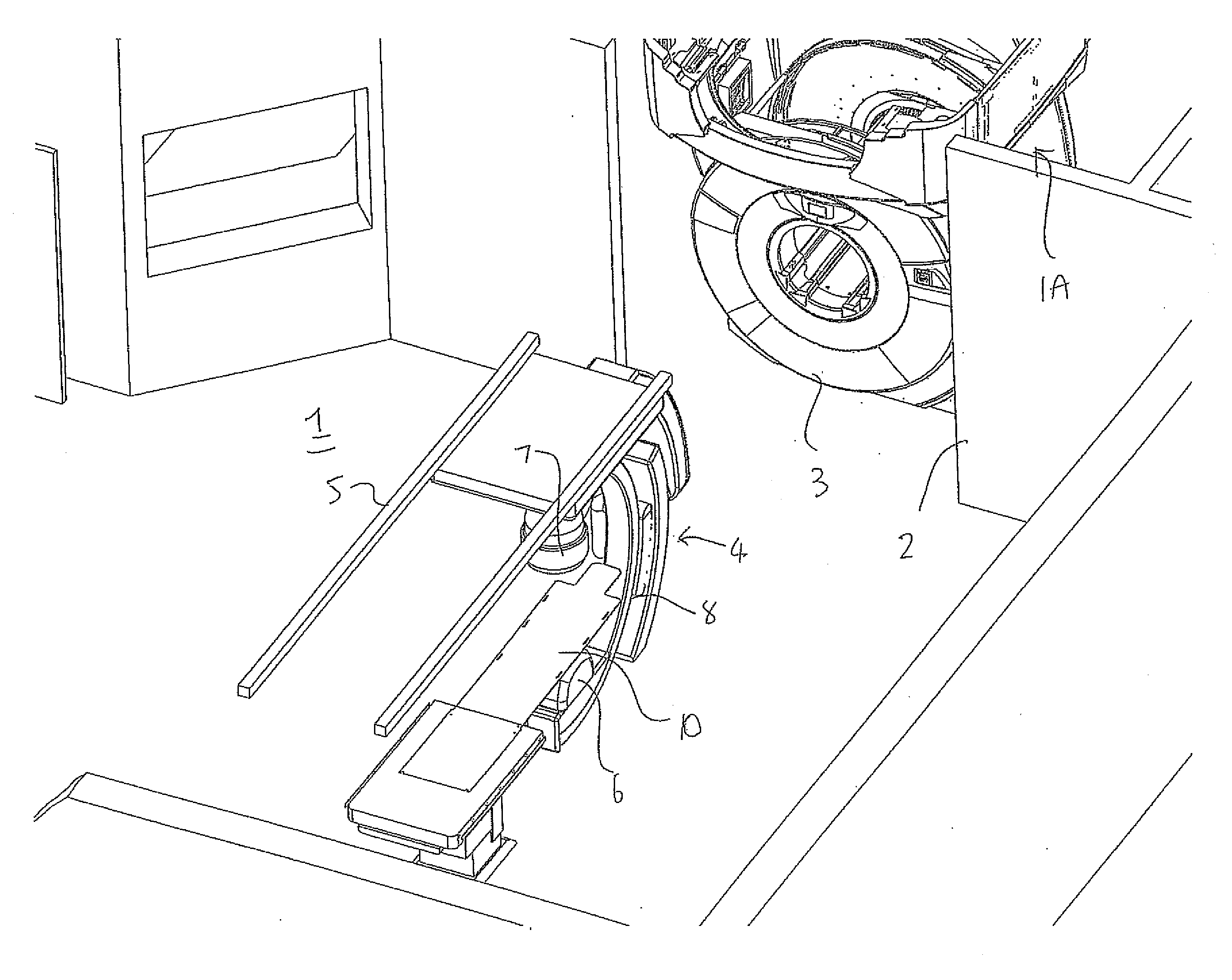
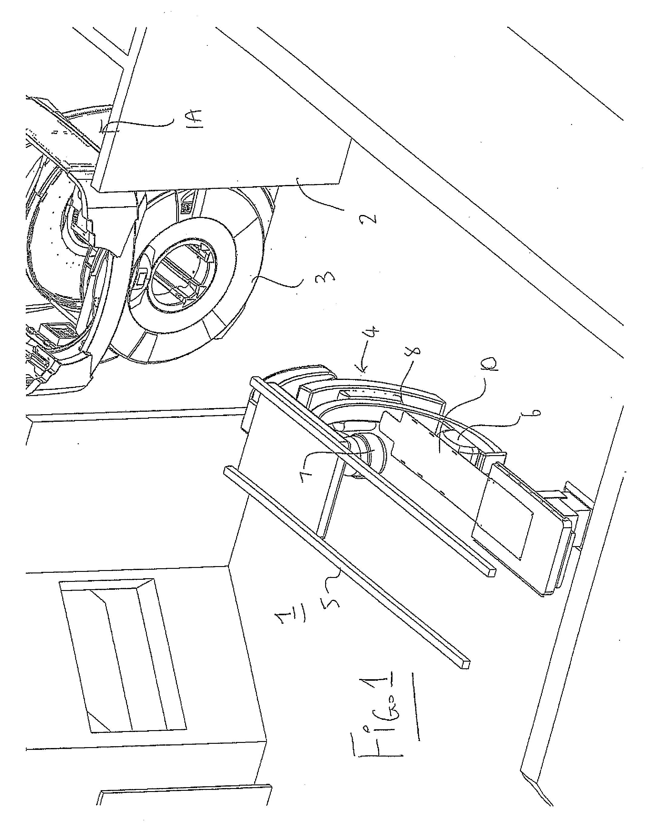
[0119]In FIG. 1 is shown an arrangement for carrying out Magnetic Resonance Imaging and X-Ray imaging of a patient while the patient remains stationary on a patient support table. The arrangement provides a room 1 in which is mounted a patient support table 10 with doors 2 at one side of the room for entry into the room of the magnet 3 of an MR imaging system from a magnet bay 1A. The room contains an X-Ray imaging system 4 mounted on rails 5 and includes an X-Ray transmitter 6 and receiver 7 mounted on a C-shaped support 8. The X-Ray system is of a conventional construction comm...


PUM


Abstract

A patient table for a common imaging system including Magnetic Resonance and X-Ray retains the patient stationary in position prior to, during and subsequent to the imaging and includes a base, a patient support portion cantilevered from the base and a mattress. A safety system is provided for controlling the operation of the magnet and MR system and the X-Ray systems to allow effective safe operation and controls the movement of the magnet to the table and the movement of the X-Ray imaging systems to and from the table to locations where they do not interfere with the MR imaging.

Description

[0001]This application claims the benefit under 35 U.S.C. 119 of Provisional Application No. 61 / 058,657 filed Jun. 4, 2008, the disclosure of which is incorporated herein by reference.[0002]This application is a continuation-in-part of application Ser. No. 12 / 333,032 filed 11 Dec. 2008.[0003]This invention relates to system for use in magnetic resonance and X-Ray imaging of a patient.BACKGROUND OF THE INVENTION[0004]With MRI, a high field magnet, typically superconducting, is arranged in a torus configuration (like a donut) and with the patient lying down inside the magnet on a table where the magnetic field allows a pulsed and sequenced magnetic and EM field to probe the body to produce images, which allow the trained radiologist to determine with high probability the anatomy of the patient. MRI is sometimes performed using contrast agents introduced to the patient to provide even better contrast between different tissue types. MRI techniques are very good at detecting the anatomic...

Claims


Application Information

Patent Timeline
IPC IPC(8): A61B5/055G01N23/04
CPCA61B5/0555A61B6/4417A61B6/0464A61B6/10A61B6/105A61B6/4441A61B6/4464A61B6/4476G01R33/34046G01R33/34084G01R33/341G01R33/381G01R33/4812A61B6/4423A61B6/0457A61B5/0035A61B6/0487A61B5/055
Inventor SCARTH, GORDONROBBINS, STEVENGUYOT, BRENDAN
Owner DEERFIELD IMAGING INC
Why Eureka
Try Eureka