Detection and localization of vascular occlusion from angiography data

a technology of angiography and localization, applied in image data processing, medical science, instruments, etc., can solve the problems of time-consuming whole procedure of iso-surface segmentation and surface display

Inactive Publication Date: 2009-12-31
AGENCY FOR SCI TECH & RES
View PDF2 Cites 91 Cited by
  • Summary
  • Abstract
  • Description
  • Claims
  • Application Information

AI Technical Summary

Benefits of technology

[0016]Preferred embodiments of the invention make it possible to detect and localise an occlusion without segmentation of the vascular data. This processing is much faster than the extraction of iso-surface and displaying it by means of t

Problems solved by technology

The whole procedure of iso-surface segmentation and surface disp

Method used


Image

  • Detection and localization of vascular occlusion from angiography data
  • Detection and localization of vascular occlusion from angiography data
  • Detection and localization of vascular occlusion from angiography data

Examples


Embodiment Construction

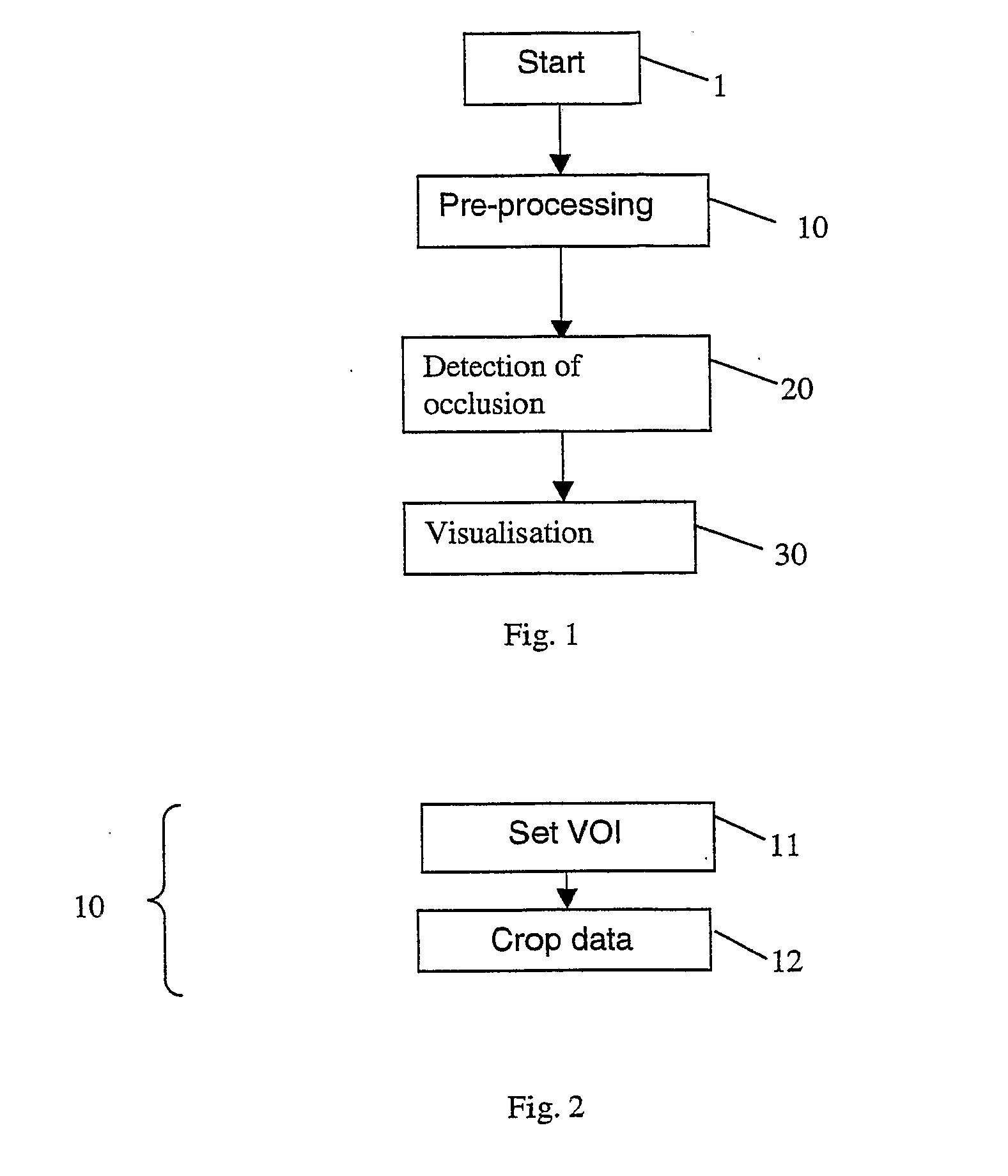
[0026]An embodiment of the invention will now be described with reference to FIG. 1. The embodiment takes as its input, at step 1, a set of three-dimensional volumetric angiographic data, which has been acquired with any suitable imaging technique, such as magnetic resonance angiography (MRA), computed tomography angiography (CTA) or X-ray rotation angiography

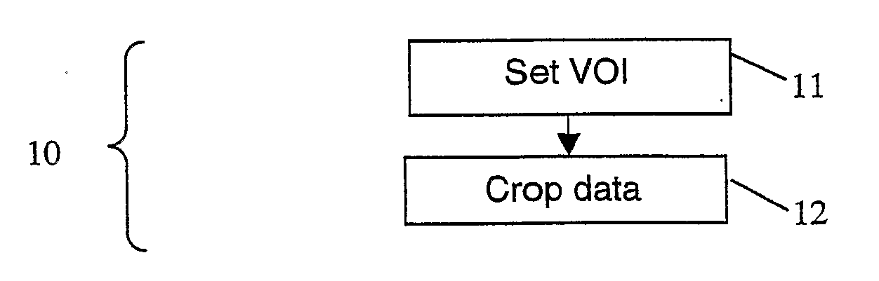
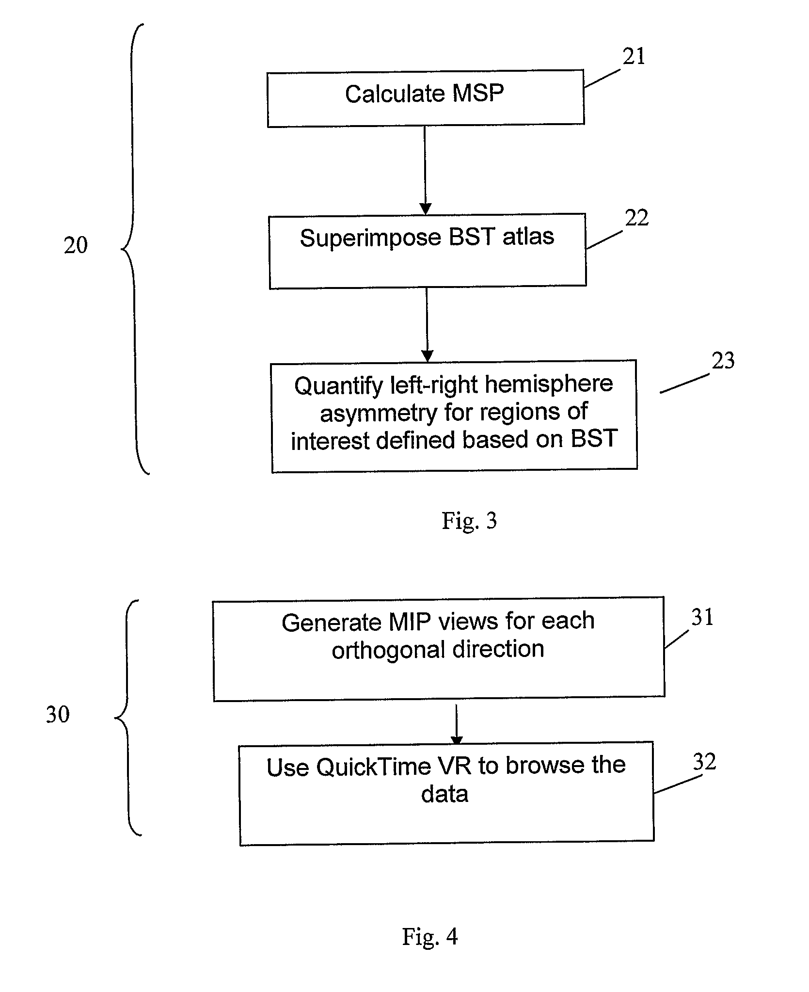
[0027]In step 10, the method pre-processes the volumetric angiographic data prior to their analysis and visualization. The sub-steps of the pre-processing step 10 are shown in FIG. 2.

[0028]The volumetric angiographic data typically depicts not only the vessels but also other structures including scalp, skull, muscles, fat, and some other bones. To facilitate analysis, some unnecessary structures are eliminated by setting a suitable volume of interest (VOI) (sub-step 11), and removing the voxels outside this volume (“cropping”—sub-step 12). The VOI can be set in several ways, depending on the specific application. In particular,...


PUM


Abstract

A technique for detecting and localising vascular occlusions in the brain of a patient is presented. The technique uses volumetric angiographic data of the brain. A mid-sagittal plane and/or lines is/are identified within the set of angiographic data. Optionally, the asymmetry of the hemispheres is measured, thereby obtaining an initial indication of whether an occlusion might be present. The angiographic data is mapped to pre-existing atlas of blood supply territories, thereby obtaining the portion of the angiographic data corresponding to each of the blood supply territories. For each territory (including any sub-territories), the asymmetry of the corresponding portion of the angiographic data about the mid-sagittal plane/lines is measured, thereby detecting any of the blood supply territory including an occlusion. The angiographic data for any such territory is displayed by a three-dimensional imaging technique.

Description

FIELD OF THE INVENTION[0001]The present invention relates to methods and computer systems for detecting whether a patient suffers from an occluded blood vessel, and in the case that occlusion is detected, to localization of the occlusion.BACKGROUND OF THE INVENTION[0002]Determination whether a blood vessel is occluded (i.e. blocked) or patent (i.e. open), and, in the case of an occlusion being identified, localization of this occlusion, are important in the diagnosis and treatment of vascular diseases. This is also critical in acute stroke management. Imaging is a means to obtain this information. Volumetric angiographic data (that is, a set of data in which elements of the data correspond to respective points within a three-dimensional region) can be acquired with various imaging techniques, such as magnetic resonance angiography (MRA), computed tomography angiography (CTA) or X-ray rotation angiography. Present techniques for detecting occlusions involve a requirement to segment v...

Claims


Application Information

Patent Timeline
IPC IPC(8): G06K9/00
CPCA61B6/504G06T7/0014G06T2207/30104G06T2207/10072G06T7/608Y10S128/922G06T7/68
Inventor NOWINSKI, WIESLAW L.
Owner AGENCY FOR SCI TECH & RES
Why Eureka
Try Eureka