Methods for differentially detecting a multimeric form from a monomeric form of a multimer-forming polypeptide through three-dimensional interactions

a multimeric form and monomeric technology, applied in the field of three-dimensional interactions for differential detection of multimeric forms from monomeric forms of multimer-forming polypeptides, can solve problems such as difficult analysis of the conformational transition that leads to the formation of prpsup>res /sup>or characterization

Active Publication Date: 2010-01-28
PEOPLEBIO
View PDF6 Cites 23 Cited by
  • Summary
  • Abstract
  • Description
  • Claims
  • Application Information

AI Technical Summary

Problems solved by technology

However, the multimeric forms often cause diseases or disorders in some proteins.
However, there has not been suggested any process and approach to differentially detect multimeric (aggregating) forms from their monomeric (normal) forms.
This latter feature makes it difficult to analyze the conformational tr

Method used


Image

  • Methods for differentially detecting a multimeric form from a monomeric form of a multimer-forming polypeptide through three-dimensional interactions
  • Methods for differentially detecting a multimeric form from a monomeric form of a multimer-forming polypeptide through three-dimensional interactions
  • Methods for differentially detecting a multimeric form from a monomeric form of a multimer-forming polypeptide through three-dimensional interactions

Examples


example i

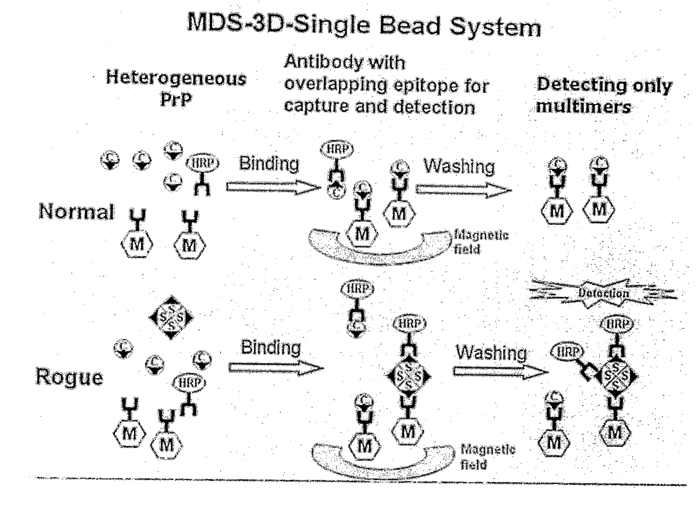
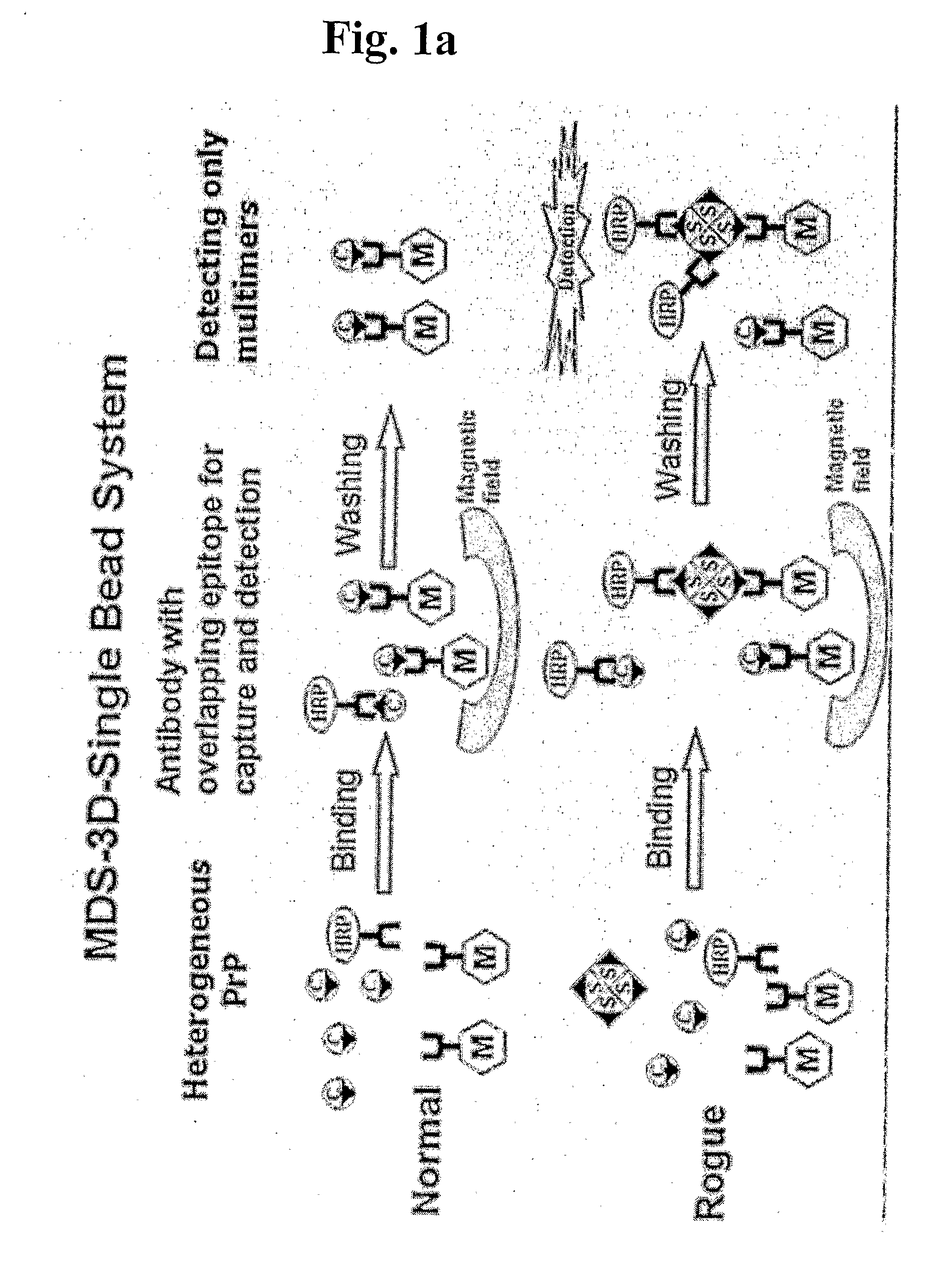
Detection of Multimeric PrP in Plasma Using MDS-3D-Sïngle Bead System

[0083]2705 μl of sheep plasma, 22.5 μl of recombinant multimeric sheep PrP (genotype ARQ, 120-fold diluted) and 3605 μl of 2.5× detergent (including 3% Triton X-IOO, 1.5% Na deoxycholate and 0.25% sarkosyl) were mixed to prepare a sample. The negative control was also prepared to contain 22.5 μl of PBS instead of the recombinant multimeric sheep PrP. Capturing antibody-conjugated magnetic beads were prepared in which 1 μg of capturing antibodies was bound to 2.5 μl of magnetic beads. The capturing antibody bound to magnetic beads is 3E7 or MA1-750 monoclonal antibodies. The epitope against the 3E7 monoclonal antibody is an amino acid sequence spanning 140-160 of PrPc (with reference to a bovine prion sequence) or 132-152 (with reference to a sheep prion sequence), which is found to be a non-repeated sequence in prion. The MA1-750 antibody recognizes specifically an epitope on PrPc, Ser-Arg-Pro-Leu-Ile-His-Phe-Gly-S...

example ii

Determination of Buffer Type Suitable in MDS-3D-Sïngle Bead System

[0089]To determine a suitable buffer type in the MDS-3D-Single Bead System of this invention, the magnetic-bound capturing antibody, 3E7-bead and detection antibody, T2-HRP were used at a weight ratio of 1 to 1 (0.8 μg:0.8 μg, substantially correspond to a mole ratio of 1 to 1) for performing the MDS-3D-Single Bead System. Plasma samples were prepared as follows: 120 μl of 10% Triton X-100 in d-H2O, 580 μl of buffer (TAPS, TBST or Tricine, pH 8.0) and 300 μl of sheep plasma showing clinical signs to have PrPsc were mixed to give 1 ml of the total volume of plasma sample containing 1.2% Triton X-100 and 30% plasma. 2 μl (0.8 μg) of 3E7-conjugated magnetic bead as capturing antibodies and 200 μl (0.8 μg) of T2-HRP (4 μg / ml in TBST) as detection antibodies were mixed to prepare a mixed antibody. 200 μl of the mixed antibody were added to 1 ml of the plasma sample and incubated for 1 hr at 37° C. The magnetic field was th...

example iii

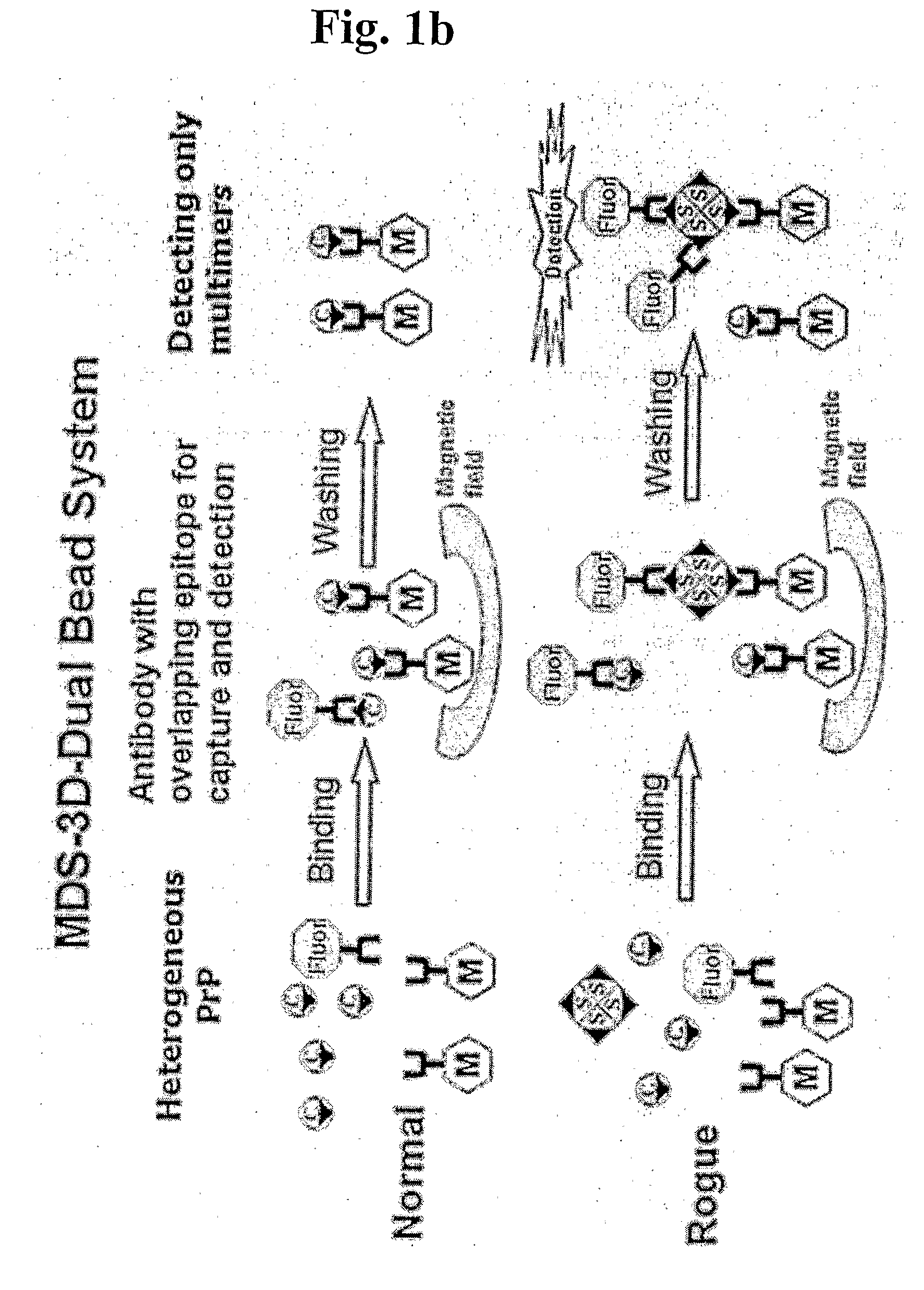
Detection of Multimeric PrP in Plasma Using MDS-3D-Dual Bead System

[0090]120 μl of 10% Triton X-100, 730 μl of TBST buffer (pH 8.0) and 150 μl of sheep plasma were mixed to give 1 ml of plasma sample containing 1.2% Triton X-100 and 15% plasma. 4 μl of 3E7-conjugated magnetic bead as capturing antibodies, 2 μl of MAI 750-conjugated fluorescence latex bead as detection antibodies and 200 μl of TBST buffer were mixed to prepare a mixed antibody (3E7-bead:MAI 750-fluorescence bead, 2:1). 200 μl of the mixed antibody were added to 1 ml of the plasma sample and incubated for 1 hr at 37° C. The magnetic field was then applied to the reaction mixture to separate magnetic beads, followed by washing the beads three times with TBST. Finally, the fluorescent signal was detected. As represented in FIG. 4, the MDS-3D-Dual Bead System permits to detect differentially PrPSc in plasma.


PUM


Abstract

The present invention relates to a method for differentially detecting a multimeric form from a monomeric form of a multimer-forming polypeptide in a biosample, which comprises the steps of: (a) preparing a carrier-capturing antibody conjugate by binding a capturing antibody to the surface of a solid phase carrier in a three dimensional manner, wherein the capturing antibody is capable of recognizing an epitope on the multimer-forming polypeptide; (b) preparing a detection antibody, wherein an epitope recognized by the detection antibody is present at a position in the multimer-forming polypeptide to cause a steric hindrance by the capturing antibody bound to its epitope to prevent the binding of the detection antibody to the multimer-forming polypeptide; (c) contacting simultaneously the carrier-capturing antibody conjugate and the detection antibody to the biosample; and (d) detecting the formation of a carrier-capturing antibody-multimeric form-detection antibody complex.

Description

BACKGROUND OF THE INVENTION[0001]1. Field of the Invention[0002]The present invention relates to methods for differentially detecting a multimeric form from a monomeric form of a multimer-forming polypeptide through three-dimensional interactions and immunoassay kits therefor.[0003]2. Description of the Related Art[0004]A multimerization of polypeptides constituting proteins has been generally known to be required for the function of proteins. However, the multimeric forms often cause diseases or disorders in some proteins. In particular, a protein exists as a monomer in normal conditions and is converted to a multimer (or aggregate form) in abnormal conditions {e.g., by the conversion to a misfolding form).[0005]It has been well established that proteins that are misfolded and ultimately aggregated (or accumulated), i.e., that are not in their functionally relevant conformation are devoid of normal biological activity. The failure to fold correctly, or to remain correctly folded, g...

Claims


Application Information

Patent Timeline
IPC IPC(8): G01N33/53G01N33/543G01N33/566G01N33/553G01N33/545
CPCG01N33/6854G01N33/54306G01N33/48G01N33/50G01N33/53
Inventor AN, SEONG SOO ALEXANDERLIM, KUN TAEKOH, HYUN JUNG
Owner PEOPLEBIO
Why Eureka
Try Eureka