
Ultrasonic bath to increase tissue perfusion

Active Publication Date: 2010-03-11
CEDARS SINAI MEDICAL CENT +1
View PDF71 Cites 2 Cited by
  • Summary
  • Abstract
  • Description
  • Claims
  • Application Information

AI Technical Summary

Benefits of technology

[0012]The present invention also describes an ultrasonic bath for enhancing tissue perfusion, enhancing development of collateral blood vessels and/or enhancing collateral circulation in an extremity in need thereof, comprising: a basin adapted to hold media and to permit the immersion of an extremity; and one or more ultrasonic transducers to generate and transmit ultrasonic energy to the subject's extremity. The basin may be about 20 inches in length, about 12 inches in width and about 40 inches in depth. In various embodiments, the one or more ultrasonic transducers may be configured to generate and transmit ultrasonic energy at a frequency of about 18 kHz to about 3 mHz. In particular embodiments, the one or more ultrasonic transducers may be configured to generate and transmit ultrasonic energy at a frequency of about 19 k

Problems solved by technology

The acute or chronic ischemia may result in pain in the extremity (e.g., claudication), loss of sensation in the extremity, sores, wounds or ulcers that heal slowly or not at all, and tissue loss in the affected extremity.
However, in many patients and especially in those with diabetes mellitus, the occlusive disease affects small blood vessels that are not amenable to mechanical revascularization.
Current technology tends to have limited long term success with high recurrence rates.
Medical therapy is also available; however, the benefit is limited.

Method used


Image

  • Ultrasonic bath to increase tissue perfusion
  • Ultrasonic bath to increase tissue perfusion
  • Ultrasonic bath to increase tissue perfusion

Examples


example 1

Transcutaneous Application of Ultrasound and Measurement of Transcutaneous Oxygen Tension

[0035]A transcutaneous oxygen tension monitor was placed on the left extremity and the corresponding right extremity. Measurement of the transcutaneous oxygen tension is a method of evaluating blood flow in settings where tissue viability and the blood flow are of concern. If the transcutaneous oxygen tension is too low, a surgeon may decide to amputate the extremity. Since the transcutaneous oxygen tension displayed by the transcutaneous oxygen tension monitor changes very frequently, a series of measurements was recorded over a short period of time (e.g., 10 seconds, 30 seconds, 1 minute, etc.) and the mean transcutaneous oxygen tension was determined and used as it provides a better indication of the transcutaneous oxygen tension.

[0036]The transcutaneous oxygen tension in both the left extremity and the corresponding right extremity were measured prior to immersing the left extremity into the...

example 2

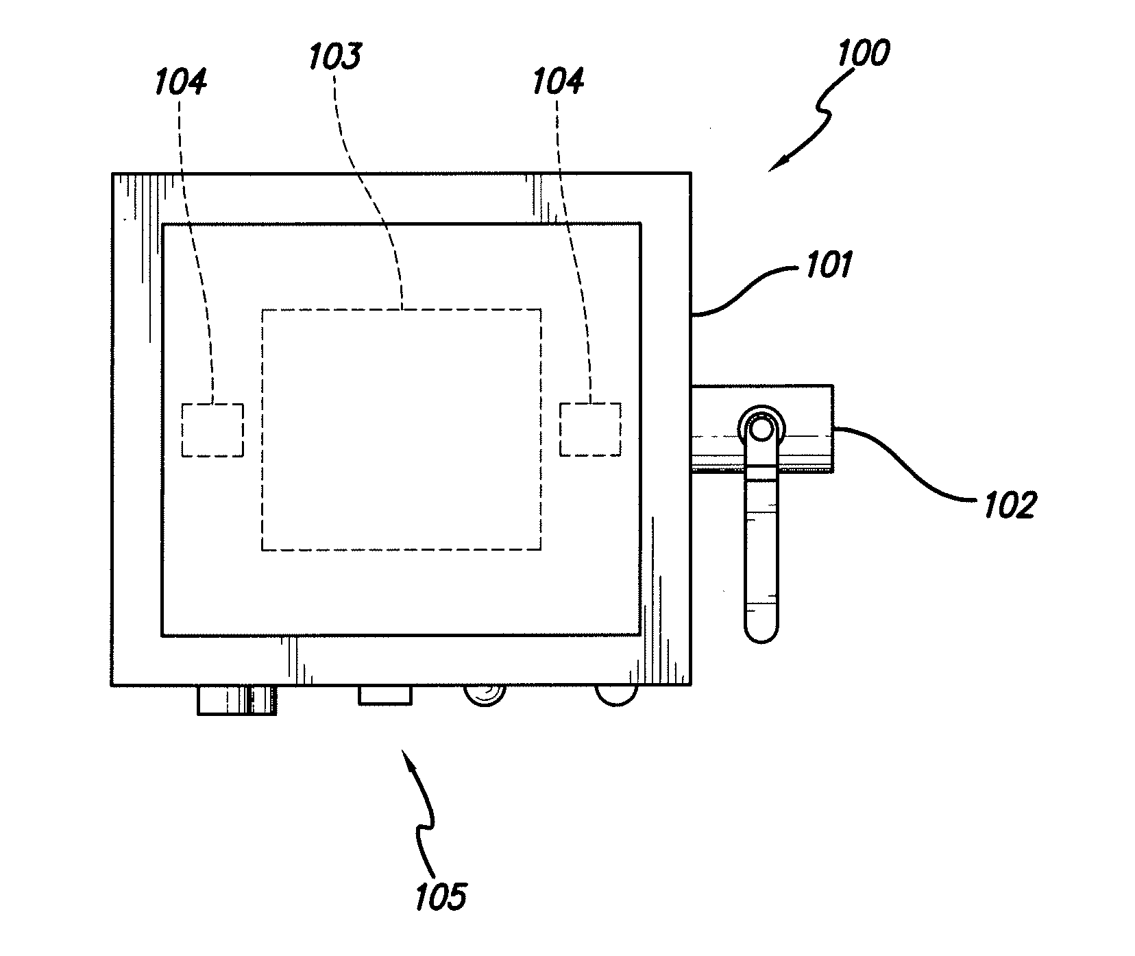
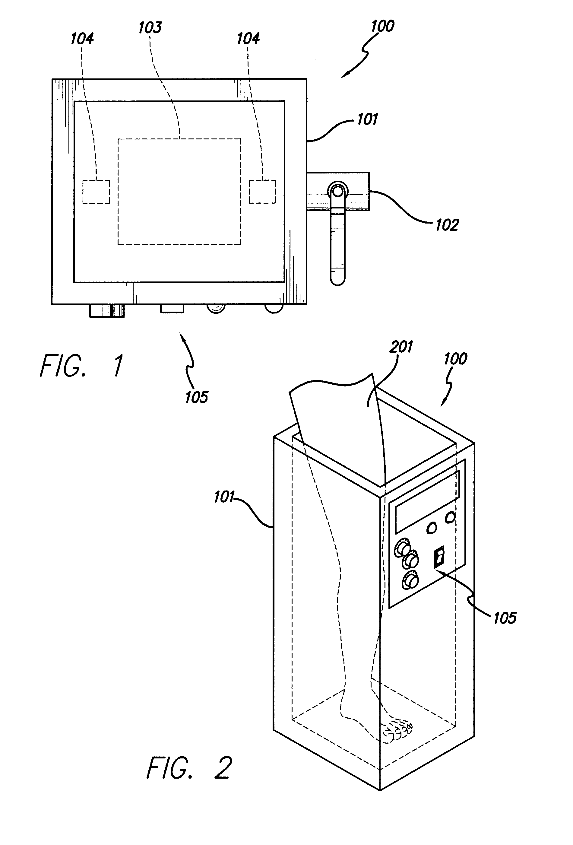
[0038]In accordance with one embodiment of the invention, FIG. 1 depicts a top view of one embodiment of the ultrasonic bath 100. As shown, the ultrasonic bath comprises a basin 101, an outlet 102, an ultrasound transducer 103 that is underneath the basin 101, heating elements 104 that are also underneath the basin 101, and a transducer control unit 105.

[0039]In accordance with another embodiment of the invention, FIG. 2 depicts a subject's leg 201 placed in the ultrasonic bath 100. The basin 101 and a transducer control unit 105 are also shown.

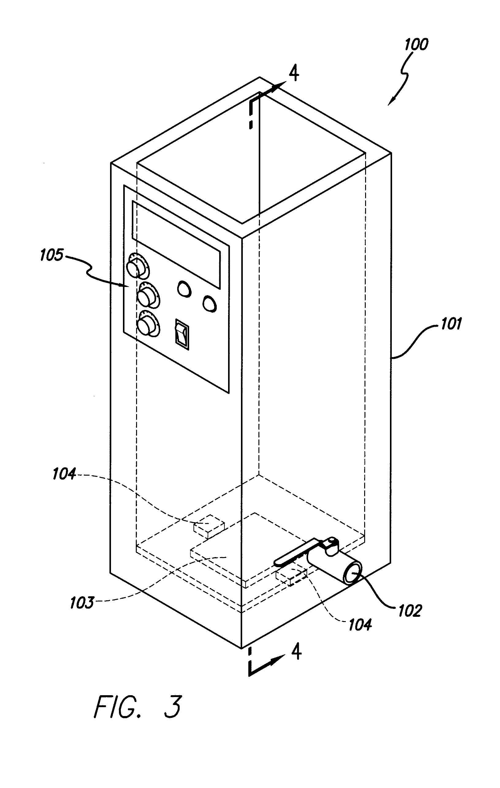
[0040]In accordance with another embodiment of the invention, FIG. 3 depicts an exterior view of an ultrasonic bath 100. An outlet 102 is used to drain the basin after use. A transducer control unit 105 is used to control the amplitude, frequency, power and / or duration of ultrasonic energy. Also shown is the transducer 103 underneath the basin 101, as well as heating elements 104 being underneath the basin 101.

[0041]In accordance with another...


PUM


Abstract

The present invention describes a method of using an ultrasonic bath to enhance tissue perfusion, enhance the development of collateral blood vessels and / or enhance collateral circulation in an extremity in need thereof. Also described is an ultrasonic bath adapted to hold media and to permit the immersion of an extremity and comprising one or more ultrasonic transducers to generate and transmit ultrasonic energy to the extremity or the inventive method

Description

FIELD OF INVENTION[0001]This invention relates to a method for using an ultrasonic bath to apply therapeutic ultrasonic energy to enhance tissue perfusion and collateral circulation and an apparatus for the same.BACKGROUND[0002]All publications herein are incorporated by reference to the same extent as if each individual publication or patent application was specifically and individually indicated to be incorporated by reference. The following description includes information that may be useful in understanding the present invention. It is not an admission that any of the information provided herein is prior art or relevant to the presently claimed invention, or that any publication specifically or implicitly referenced is prior art.[0003]Peripheral Vascular Disease (“PVD”), also referred to as Peripheral Arterial Disease (“PAD”), is a general term for diseases caused by occlusion or obstruction of peripheral arteries. PVD is a common disease affecting numerous patients that may lea...

Claims


Application Information

Patent Timeline
IPC IPC(8): A61N7/00
CPCA61N7/00A61H35/00A61H33/00A61N2007/0004
Inventor SIEGEL, ROBERT J.BIRNBAUM, YOCHAILUO, HUAI
Owner CEDARS SINAI MEDICAL CENT
Learn More