Decellularization and recellularization apparatuses and systems containing the same

a technology of recellularization apparatus and recellularization composition, which is applied in the direction of biochemical apparatus and processes, specific use bioreactors/fermenters, prosthesis, etc., can solve the problems of the successful recellularization of the scaffold, compromising the quality and integrity of the scaffold or its constituents, and affecting the use of the scaffold. , to achieve the effect of reducing and minimizing the residency of the decellularization composition

Inactive Publication Date: 2010-04-15
MIROMATRIX MEDICAL
View PDF22 Cites 38 Cited by
  • Summary
  • Abstract
  • Description
  • Claims
  • Application Information

AI Technical Summary

Benefits of technology

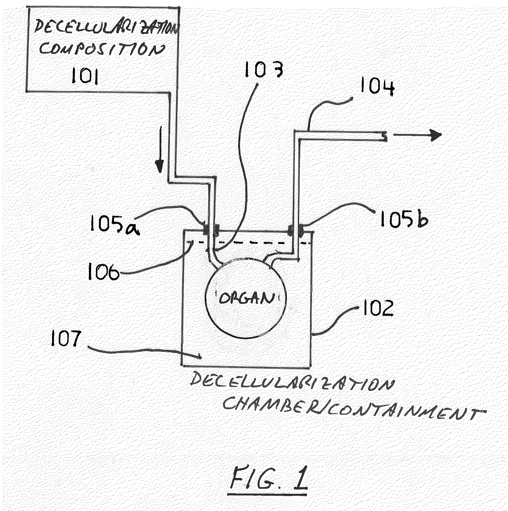
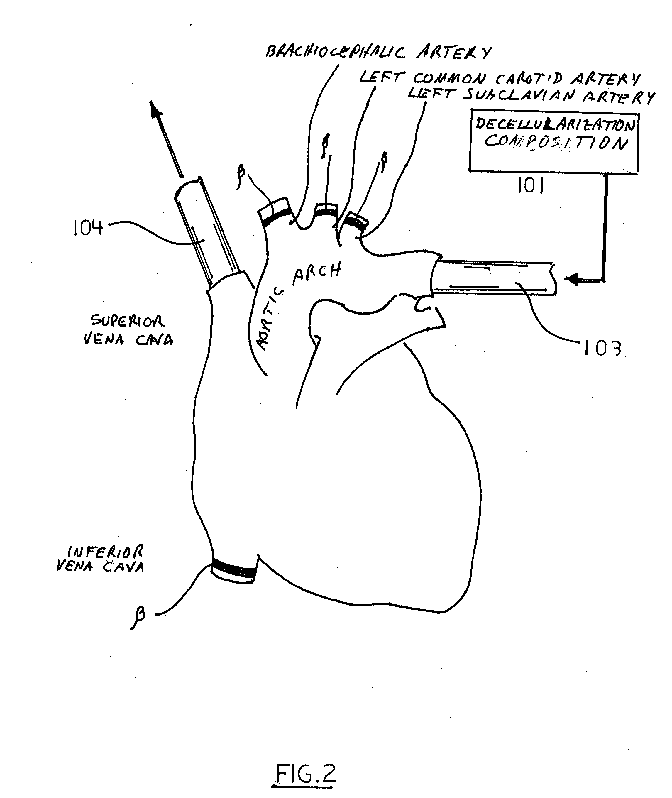
[0006]The invention provides a decellularization apparatus comprising: a decellularization chamber; a decellularization composition reservoir; an organ / tissue ingress conduit connected to the decellularization composition reservoir for delivery of the composition into the organ or tissue, the ingress conduit comprising a natural anatomical conduit engagement structure (e.g., vascular or duct engagement structure); and a organ / tissue egress conduit comprising a natural anatomical conduit engagement structure (e.g., vascular engagement structure). The decellularization apparatus can further comprise an organ / tissue positioning structure. The decellularization apparatus is structured to 1) cooperate with and utilize the natural anatomical conduits (e.g., natural vasculature or duct) of the target organ or tissue for delivery of a decellularization composition throughout the organ or tissue; and 2) reduce and minimize residency of the decellularization composition associated with the organ or tissue. The decellularization apparatus preferably maintains an aseptic environment throughout the apparatus components.
[0007]The invention also provides a decellularization system comprising a decellularization apparatus as described above in combination with a decellularization composition. The combined features of the decellularization system effectuate detachment and separation of the cellular material from the target organ or tissue to produce the extracellular matrix-based scaffold without the need for, mechanical disruption techniques.

Problems solved by technology

Currently used techniques and equipment for decellularizing organs or tissues can substantially damage the extracellular matrix and membranous tissues associated with a given organ or tissue both physically and biochemically.
This in turn can compromise the quality and integrity of the scaffold or its constituents, thereby adversely affecting the use of the scaffold and successful recellularization of the scaffold.
As a further disadvantage, current techniques and equipment can also compromise and attenuate desirable biochemical attributes that were present in the intact natural initial extracellular componentry of the organ or tissue.
The disadvantage associated with these methods is significant compromise of the original intact scaffold in terms of both physical and chemical properties.

Method used


Image

  • Decellularization and recellularization apparatuses and systems containing the same
  • Decellularization and recellularization apparatuses and systems containing the same
  • Decellularization and recellularization apparatuses and systems containing the same

Examples


example

Example 1

Comparative Data: Perfusion Organ Decellularization Versus Immersion Decellularization Techniques Using Kidney

[0155]Using a kidney as the organ, the organ was decellularized using the immersion methods described in U.S. Pat. Nos. 6,753,181 and 6,376,244, incorporated herein by reference. Briefly, an organ was placed in dH2O and agitated with a magnetic stir bar rotating at 100 rpm for 48 hours at 4° C., and then the organ was transferred to an 10 ammonium hydroxide (0.05%) and Triton X-100 (0.5%) solution for 48 hours with continued magnetic stir bar (100 rpm) stirring of the solution. The solution was changed and the 48 hr immersion with the ammonium hydroxide and Triton X-100 was repeated as needed to decellularize the organ (generally a visual acellular organ). The liver took approximately 5 repetitions of ammonium 15 hydroxide and Triton X-100 to generate a visually acellular organ. After the decellularization process, organs were transferred to dH2O for 48 hours with a...


PUM

PropertyMeasurementUnit
temperatureaaaaaaaaaa
body temperatureaaaaaaaaaa

Abstract

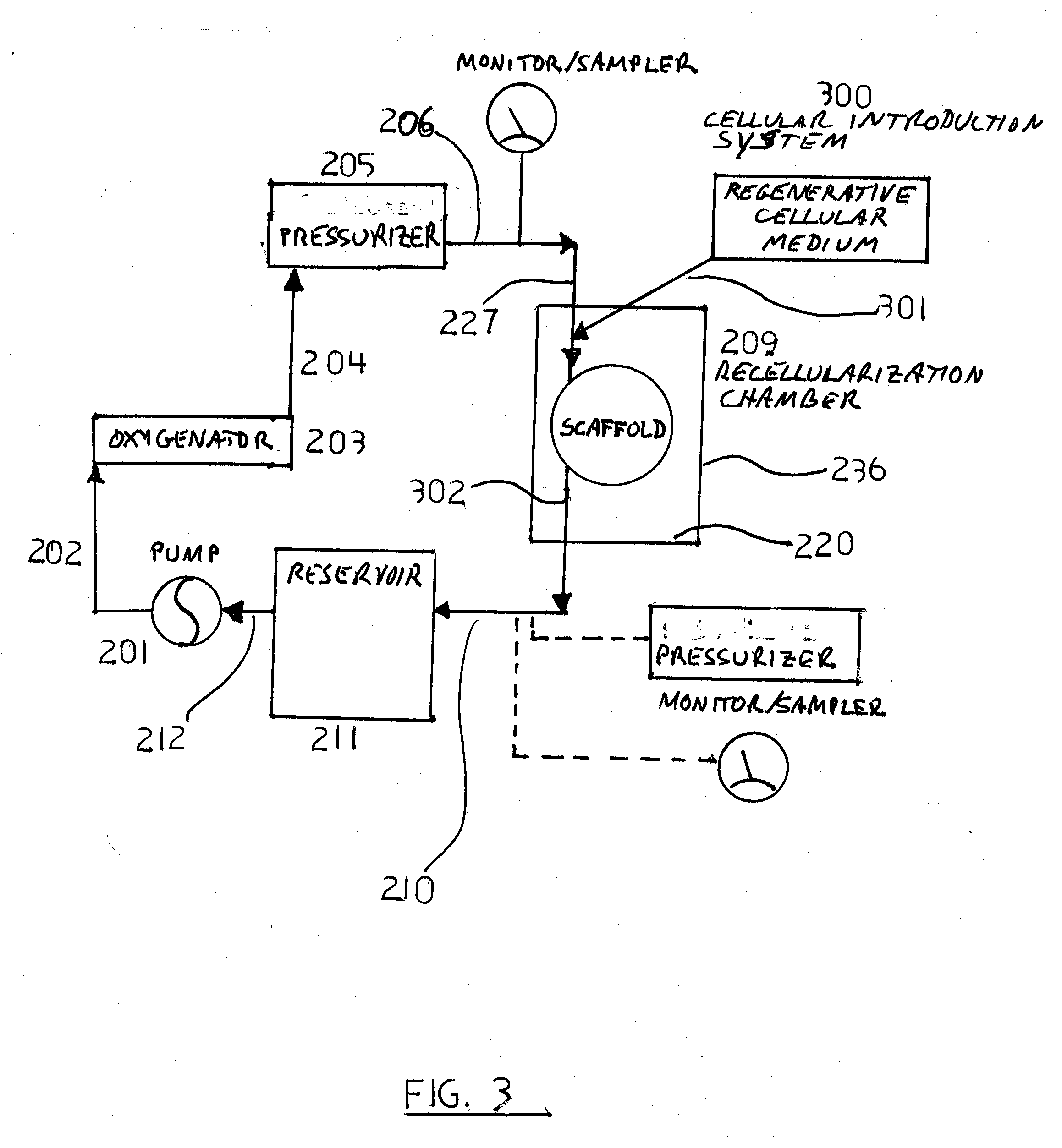
The invention described herein provides systems and apparatuses for an initial preparation of an organ or tissue scaffold comprising an extracellular matrix, and subsequent recellularization of the scaffold to ultimately form a resultant artificial organ or tissue incorporating the natural and original extracellular matrix. The techniques and equipment of the invention collectively minimize scaffold collapse, compression or physical damage to the organ as well as afford the advantages of significant maintenance of the initial natural structural and biochemical attributes of the organ or tissue. The invention is particularly useful in organ and tissue transplantation and repair.

Description

RELATED APPLICATION DATA[0001]This application is a continuation-in-part of, and claims benefit under 35 U.S.C. §120 to, pending U.S. application Ser. No. 12 / 064,613 deposited with the U.S. Patent & Trademark Office on Feb. 22, 2008, which claims benefit under 35 U.S.C. §371 to International Application No. PCT / US2006 / 033415 filed Aug. 28, 2006, which claims benefit under 35 U.S.C: §119(e) to U.S. Provisional Application Nos. 60 / 711,501 filed Aug. 26, 2005 and 60 / 815,242 filed Jun. 19, 2006. This application also claims benefit under 35 U.S.C. §119(e) to U.S. Provisional Application No. 61 / 211,613 filed Mar. 31, 2009.BACKGROUND OF THE INVENTION[0002]Biologically derived matrices have been developed for tissue engineering and regeneration and are known. Matrices and scaffolds developed to date, however, generally have numerous disadvantages associated with them. Currently used techniques and equipment for decellularizing organs or tissues can substantially damage the extracellular ma...

Claims


Application Information

Patent Timeline
Patent Type & Authority Applications(United States)
IPC IPC(8): C12M3/00A61K35/407A61K35/55
CPCA61K35/407C12M41/40A61L27/3683A61L27/38A61L27/54A61L2300/254A61L2300/414A61L2300/42A61L2300/426A61L2300/428A61L2300/43A61L2300/434A61L2430/40C12M21/08C12M25/14C12M29/10A61K35/55
Inventor TAYLOR, DORIS A.KREN, STEFAN M.ROBERTSON, MATTHEW JEFFREY
Owner MIROMATRIX MEDICAL
Why Eureka
Try Eureka