Learning anatomy dependent viewing parameters on medical viewing workstations

a technology of viewing workstations and viewing parameters, which is applied in the field of data processing apparatuses providing visualisation parameters controlling the display of medical images, can solve the problems of difficult comparison or analysis of medical images, misinterpretation of medical images, etc., and achieves the improvement of pathology monitoring, simplified viewing of medical data sets, and improved inter-patient comparison of images.

Inactive Publication Date: 2010-04-22
KONINKLIJKE PHILIPS ELECTRONICS NV
View PDF8 Cites 14 Cited by
  • Summary
  • Abstract
  • Description
  • Claims
  • Application Information

AI Technical Summary

Benefits of technology

[0012]The data processing apparatus described above may provide a new functionality for medical viewing workstations: after learning a certain anatomical view (e.g. four chamber view of the human heart), the user can automatically “jump” to these views and display the data in fixed anatomy dependent perspective, using learned contrast or lighting parameters. This new feature will not only simplify the viewing of medical data sets, but it will also enable a better inter-patient comparison of images (slice to slice correspondence) and it will improve the monitoring of pathologies in follow up studies. The data processing apparatus according to the invention provides fully automated adjustment of viewing, perspective, and contrast parameters on medical viewing workstations. When the above mentioned comparison module has identified a data set that is identical or sufficiently similar to the current data set, then retrieval of the visualisation parameters comprises extracting these visualisation parameters from the record containing the identified data set. In the alternative to storing both the data set and the visualisation parameters in the same record, one of them or both may contain references to each other.

Problems solved by technology

This makes a comparison or analysis of medical images difficult.
Moreover, manual adjustments may be a potential source of error leading to a misinterpretation of the medical image.

Method used


Image

  • Learning anatomy dependent viewing parameters on medical viewing workstations
  • Learning anatomy dependent viewing parameters on medical viewing workstations
  • Learning anatomy dependent viewing parameters on medical viewing workstations

Examples


Embodiment Construction

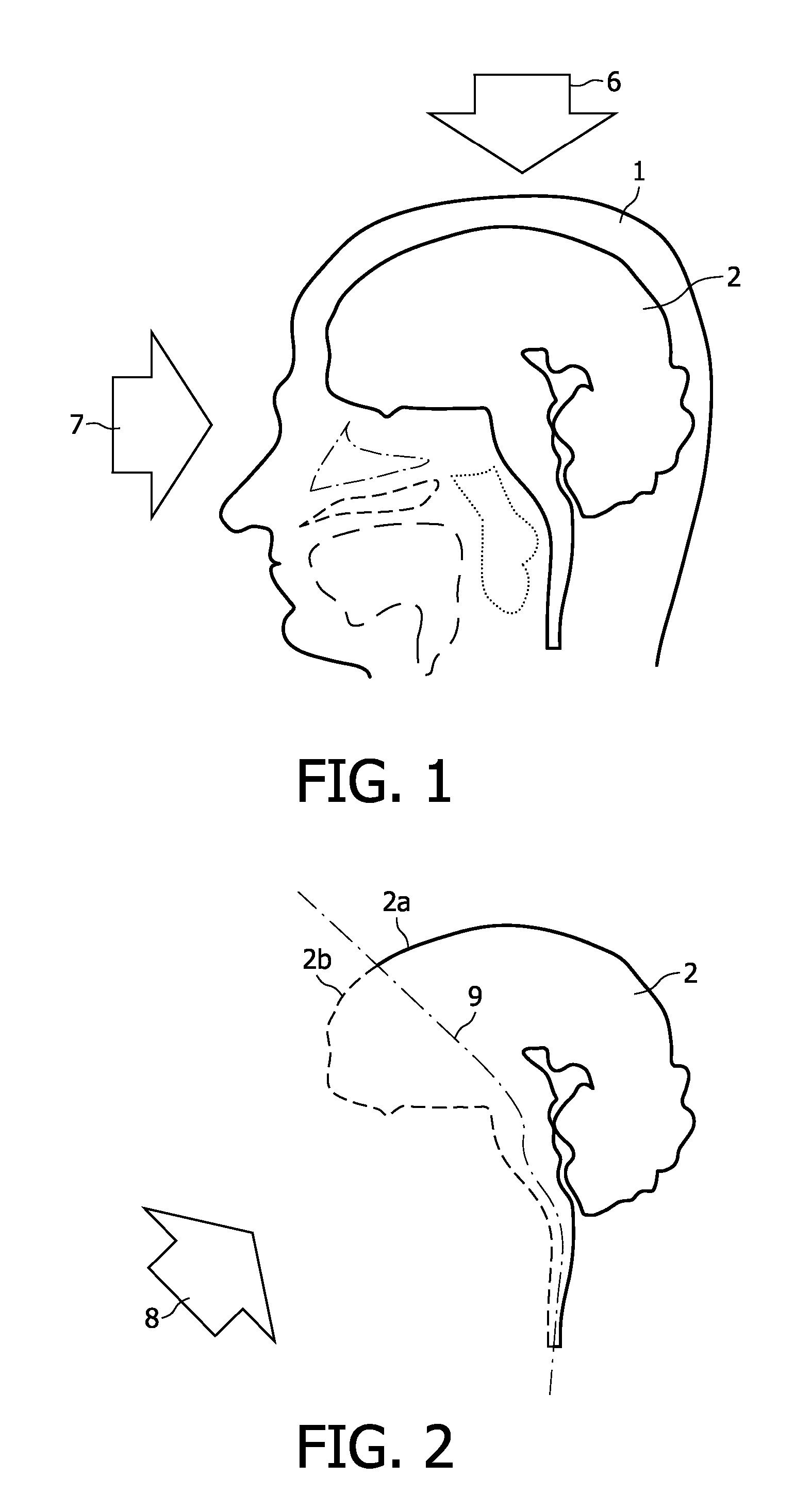
[0044]The various aspects of the present invention can be readily understood by first studying an exemplary application. FIG. 1 shows the head 1 of a human patient. The brain 2 and various other anatomical structures within the head of the patient, such as the tongue or the palate are also represented in a schematic manner. Let us assume that a user of the medical imaging modality is mainly interested in visualising the brain 2. The user may be a physician, a radiologist, or another person involved with the acquisition and visualisation of medical images. Let us further assume that a three-dimensional scan of the patient's head is available. Depending on which region of the brain 2 the user desires to exam, he needs to choose an appropriate perspective. Two of several possible perspectives are represented in FIG. 1 by the arrows 6 and 7. Arrow 6 represents a perspective in which the user looks down onto the top of the brain 2. Arrow 7 represents another perspective corresponding to ...


PUM


Abstract

The invention relates to a data processing apparatus and a method for providing visualisation parameters controlling the display of a medical image (12). The data processing apparatus comprises a mapping component (16). The mapping component (16) is arranged to receive a current dataset (15) corresponding to the medical image and comprising a content description thereof, to compare the content description of the current dataset (15) with a content description of a plurality of stored datasets, to select at least one further dataset out of the plurality of stored datasets, to retrieve stored visualisation parameters corresponding to the at least one further dataset, and to prepare the retrieved visualisation parameters as the visualisation parameters controlling the display of the medical image (12).

Description

FIELD OF THE INVENTION[0001]The invention relates to a data processing apparatus for providing visualisation parameters controlling the display of a medical image.[0002]The invention further relates to a method of providing visualisation parameters controlling the display of a medical image. The invention still further relates to a computer program product.BACKGROUND OF THE INVENTION[0003]It is known from international patent application WO 2006 / 013499 to analyze a medical image with respect to a spatial position and orientation of an object shown in the medical image. The result of the analysis is used in order to define the parameters of the scan geometry for a new scan of the same object. Typically, the new scan is performed at a higher resolution or other modified settings of the scan process compared to the first scan. As such, the first scan is often referred to as a scout scan. Automated scan planning enables fully automated acquisition of medical data sets of MRI (Magnetic R...

Claims


Application Information

Patent Timeline
Patent Type & Authority Applications(United States)
IPC IPC(8): G06F17/30G06F3/048G06F19/00
CPCG06F19/321G06T2210/41G06T19/00G16H30/20G16H30/40
Inventor BYSTROV, DANIELYOUNG, STEWARTPEKAR, VLADIMIRCOCOSCO, CHRISTIAN ADRIAN
Owner KONINKLIJKE PHILIPS ELECTRONICS NV
Why Eureka
Try Eureka