Method for producing a stereotactic image in a mammography device

a stereotactic image and mammography technology, applied in the field of stereotactic image production in the mammography device, can solve the problems of affecting the spatial accuracy of the biopsy, the complete procedure described above is therefore extremely time-consuming, etc., and achieves the effect of improving the mammography or biopsy method, significantly reducing the time duration of the whole procedur

Inactive Publication Date: 2010-04-29
SIEMENS AG
View PDF10 Cites 8 Cited by
  • Summary
  • Abstract
  • Description
  • Claims
  • Application Information

AI Technical Summary

Benefits of technology

[0010]An object of the invention is to improve the mammography or biopsy method by significantly reducing t

Problems solved by technology

This complete procedure described above is therefore extremely time-consuming.
During this entire, extremely time-consuming procedure, which is at the least uncomfortable if not actually very painful, the patient rem

Method used


Image

  • Method for producing a stereotactic image in a mammography device
  • Method for producing a stereotactic image in a mammography device

Examples


Embodiment Construction

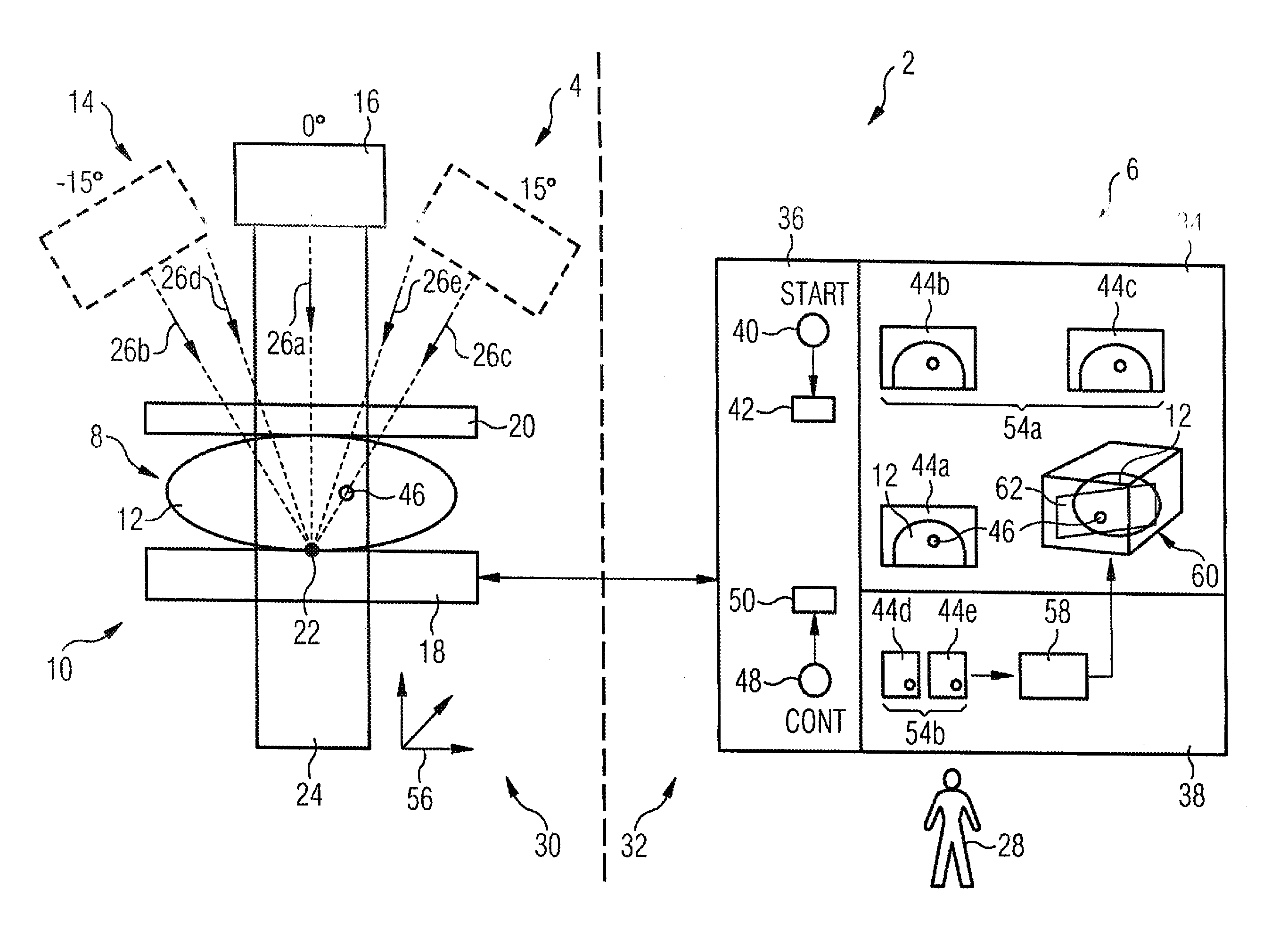
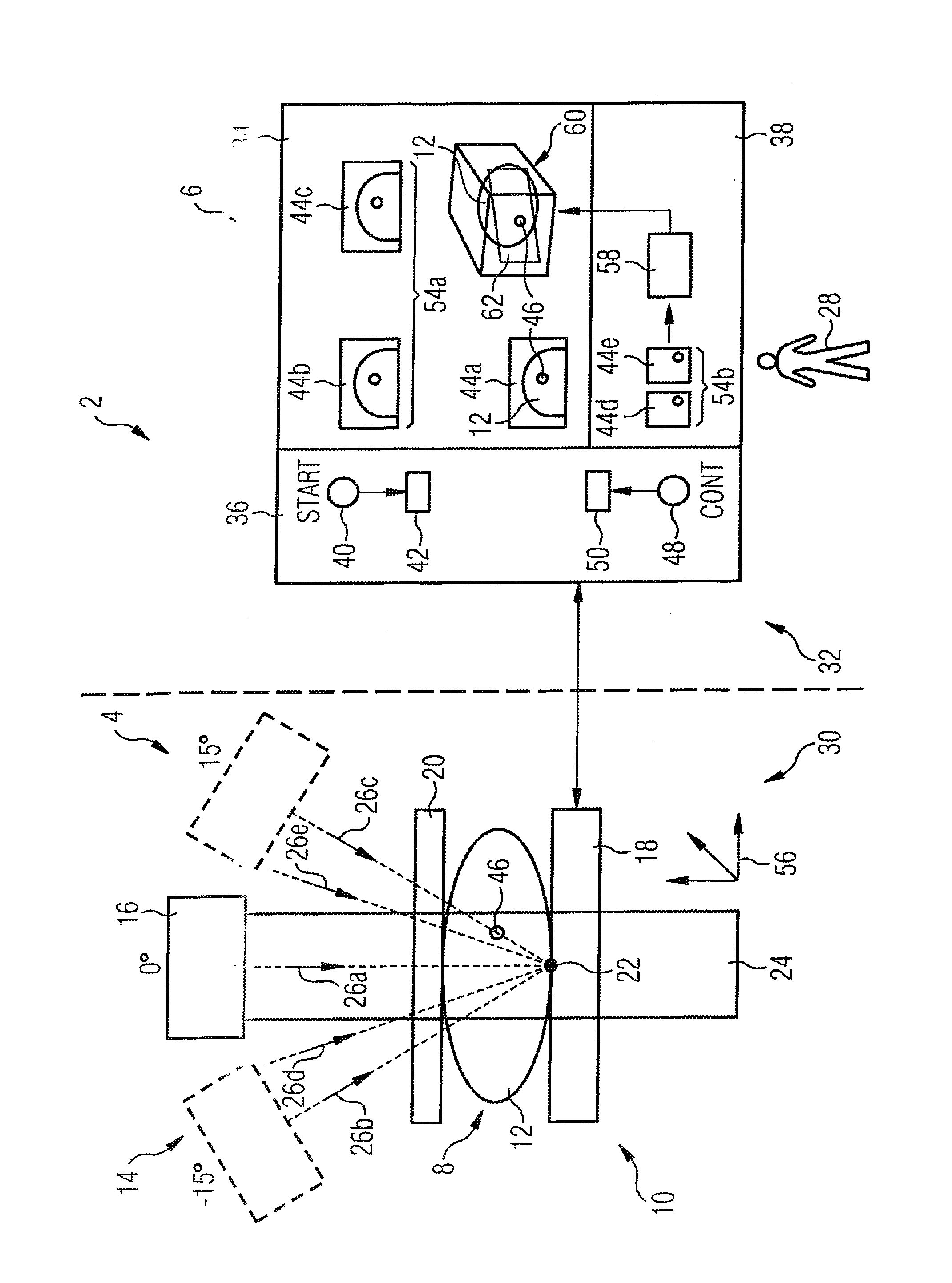
[0025]The FIGURE shows a mammography system 2 that includes a mammography apparatus 4 in a treatment room 30 and a workstation 6 in a control room 32. Treatment room 30 and control room 32 are shielded from one another in terms of radiation (indicated by a dashed line). Especially in mammography it is also typical that treatment room 30 and control room 32 are a single room, wherein both regions (i.e. mammography apparatus 4 and workstation 6) are separated by a radiation protection wall.

[0026]A patient 8 (of whom only the breast 12 held in the compression device 10 is shown) is fixed in the mammography apparatus 4, i.e. in the compression device 10 thereof. The mammography device 4 also has an x-ray device 14. The x-ray device 14 has an x-ray source 16 and a flat panel detector 18. The flat panel detector 18, together with a compression plate 20, forms the compression device 10 to fix the breast 12. The x-ray device 14 or at least the x-ray source 16 thereof is supported on a stand...


PUM


Abstract

In a method for producing a stereotactic image of a patient fixed in a mammography device for a biopsy, a computerized control and evaluation unit waits for a start signal to be given by an operator, and after receipt of the start signal, automatically implements the following steps. An x-ray apparatus of the mammography device is positioned in a first predetermined imaging position, and a first image of the patient is acquired with the x-ray apparatus in this position. The x-ray apparatus is then pivoted into a second imaging position, different from the first, and a second image of the patient is acquired at the second position. The first and second images are acquired to form a stereotactic image of the patient.

Description

BACKGROUND OF THE INVENTION[0001]1. Field of the Invention[0002]The present invention concerns a method to generate a stereotactic image of a patient fixed in a mammography system for a biopsy to be conducted on the patient.[0003]2. Description of the Prior Art[0004]Mammography systems normally serve for the x-ray examination of a breast of (typically) a female patient. If a suspicious point in the breast is diagnosed upon such an examination, a biopsy on the patient is also implemented with the aid of the mammography system, in which a biopsy needle is directed to the suspicious point in order to extract a tissue sample. In order to precisely hit the suspicious point with the biopsy needle, a localization of the suspicious point in the coordinate system of the mammography system is necessary.[0005]For an image-assisted breast biopsy (also called a stereotactic biopsy), two x-ray images or mammograms are typically produced from slightly different viewing directions in order to enabl...

Claims


Application Information

Patent Timeline
IPC IPC(8): G06K9/00
CPCA61B6/022A61B6/502A61B6/025
Inventor HALL, MICHAELHANSROUL, MARCJOHANSSON, KATRINMERTELMEIER, THOMASSAVARD, SERGE
Owner SIEMENS AG
Why Eureka
Try Eureka