Surgical access system and related methods

a surgical and access system technology, applied in the field of surgical access systems, can solve the problems of increasing health care costs, concomitant high pain, and long hospitalization, and achieve the effect of ensuring the structural stability of the retractor blad

Active Publication Date: 2010-06-17
NUVASIVE
View PDF7 Cites 126 Cited by
  • Summary
  • Abstract
  • Description
  • Claims
  • Application Information

AI Technical Summary

Benefits of technology

[0012]According to one broad aspect of the present invention, the access system comprises a tissue distraction assembly and a tissue retraction assembly, both of which may be equipped with one or more electrodes for use in detecting the existence of (and optionally the distance and / or direction to) neural structures. The tissue distraction assembly (in conjunction with one or more elements of the tissue retraction assembly) is capable of, as an initial step, distracting a region of tissue between the skin of the patient and the surgical target site. The tissue retraction assembly is capable of, as a secondary step, being introduced into this distracted region to thereby define and establish the operative corridor. Once established, any of a variety of surgical instruments, devices, or implants may be passed through and / or manipulated within the operative corridor depending upon the given surgical procedure. The electrode(s) are capable of, during both tissue distraction and retraction, detecting the existence of (and optionally the distance and / or direction to) neural structures such that the operative corridor may be established through (or near) any of a variety of tissues having such neural structures which, if contacted or impinged, may otherwise result in neural impairment for the patient. In this fashion, the access system of the present invention may be used to traverse tissue that would ordinarily be deemed unsafe or undesirable, thereby broadening the number of manners in which a given surgical target site may be accessed.
[0015]The retractor blades may be optionally dimensioned to receive and direct a rigid shim element to augment the structural stability of the retractor blades and thereby ensure the operative corridor, once established, will not decrease or become more restricted, such as may result if distal ends of the retractor blades were permitted to “slide” or otherwise move in response to the force exerted by the displaced tissue. In a preferred embodiment, only the posterior retractor blade is equipped with such a rigid shim element. In an optional aspect, this shim element may be advanced into the disc space after the posterior retractor blade is positioned, but before the retractor is opened into the fully retracted position. The rigid shim element is preferably oriented within the disc space such that is distracts the adjacent vertebral bodies, which serves to restore disc height. It also preferably advances a sufficient distance within the disc space (preferably past the midline), which serves the dual purpose of preventing post-operative scoliosis and forming a protective barrier (preventing the migration of tissue (such as nerve roots) into the operative field and the inadvertent advancement of instruments outside the operative field).
[0016]The retractor blades may optionally be equipped with a mechanism for transporting or emitting light at or near the surgical target site to aid the surgeon's ability to visualize the surgical target site, instruments and / or implants during the given surgical procedure. According to one embodiment, this mechanism may comprise, but need not be limited to, providing one or more strands of fiber optic cable within the walls of the retractor blades such that the terminal (distal) ends are capable of emitting light at or near the surgical target site. According to another embodiment, this mechanism may comprise, but need not be limited to, constructing the retractor blades of suitable material (such as clear polycarbonate) and configuration such that light may be transmitted generally distally through the walls of the retractor blade light to shine light at or near the surgical target site. This may be performed by providing the retractor blades having light-transmission characteristics (such as with clear polycarbonate construction) and transmitting the light almost entirely within the walls of the retractor blade (such as by frosting or otherwise rendering opaque portions of the exterior and / or interior) until it exits a portion along the interior (or medially-facing) surface of the retractor blade to shine at or near the surgical target site. The exit portion may be optimally configured such that the light is directed towards the approximate center of the surgical target site and may be provided along the entire inner periphery of the retractor blade or one or more portions therealong.

Problems solved by technology

Open surgical techniques are generally undesirable in that they typically require large incisions and high amounts of tissue displacement to gain access to the surgical target site, which produces concomitantly high amounts of pain, lengthened hospitalization (increasing health care costs), and high morbidity in the patient population.
The access systems developed to date, however, fail in various respects to meet all the needs of the surgeon population.
One drawback associated with prior art surgical access systems relates to the ease with which the operative corridor can be created, as well as maintained over time, depending upon the particular surgical target site.
For example, when accessing surgical target sites located beneath or behind musculature or other relatively strong tissue (such as, by way of example only, the psoas muscle adjacent to the spine), it has been found that advancing an operative corridor-establishing instrument directly through such tissues can be challenging and / or lead to unwanted or undesirable effects (such as stressing or tearing the tissues).
While certain efforts have been undertaken to reduce the trauma to tissue while creating an operative corridor, such as (by way of example only) the sequential dilation system of U.S. Pat. No. 5,792,044 to Foley et al., these attempts are nonetheless limited in their applicability based on the relatively narrow operative corridor.
More specifically, based on the generally cylindrical nature of the so-called “working cannula,” the degree to which instruments can be manipulated and / or angled within the cannula can be generally limited or restrictive, particularly if the surgical target site is a relatively deep within the patient.
While this system may provide for improved instrument manipulation relative to sequential dilation access systems (at least at deep sites within the patient), it is nonetheless flawed in that the deployment of the expandable portion may inadvertently compress or impinge upon sensitive tissues adjacent to the surgical target site.
For example, in anatomical regions having neural and / or vasculature structures, such a blind expansion may cause the expandable portion to impinge upon these sensitive tissues and cause neural and / or vasculature compromise, damage and / or pain for the patient.
This highlights yet another drawback with the prior art surgical access systems, namely, the challenges in establishing an operative corridor through or near tissue having major neural structures which, if contacted or impinged, may result in neural impairment for the patient.
Due to the threat of contacting such neural structures, efforts thus far have largely restricted to establishing operative corridors through tissue having little or substantially reduced neural structures, which effectively limits the number of ways a given surgical target site can be accessed.
However, they are nonetheless disadvantageous in that they require traversing through a much greater distance within the patient to establish the operative corridor, oftentimes requiring an additional surgeon to assist with moving the various internal organs out of the way to create the operative corridor.

Method used


Image

  • Surgical access system and related methods
  • Surgical access system and related methods
  • Surgical access system and related methods

Examples


Embodiment Construction

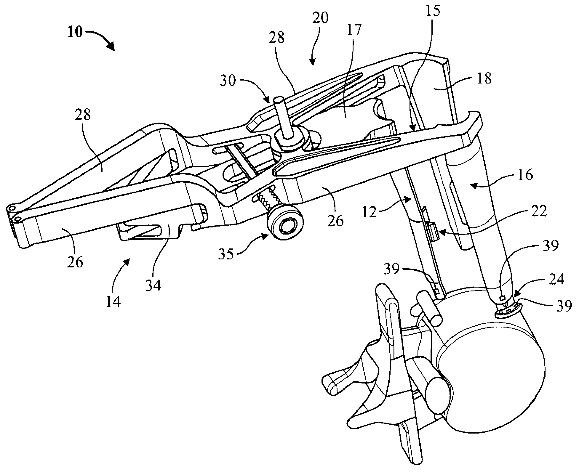
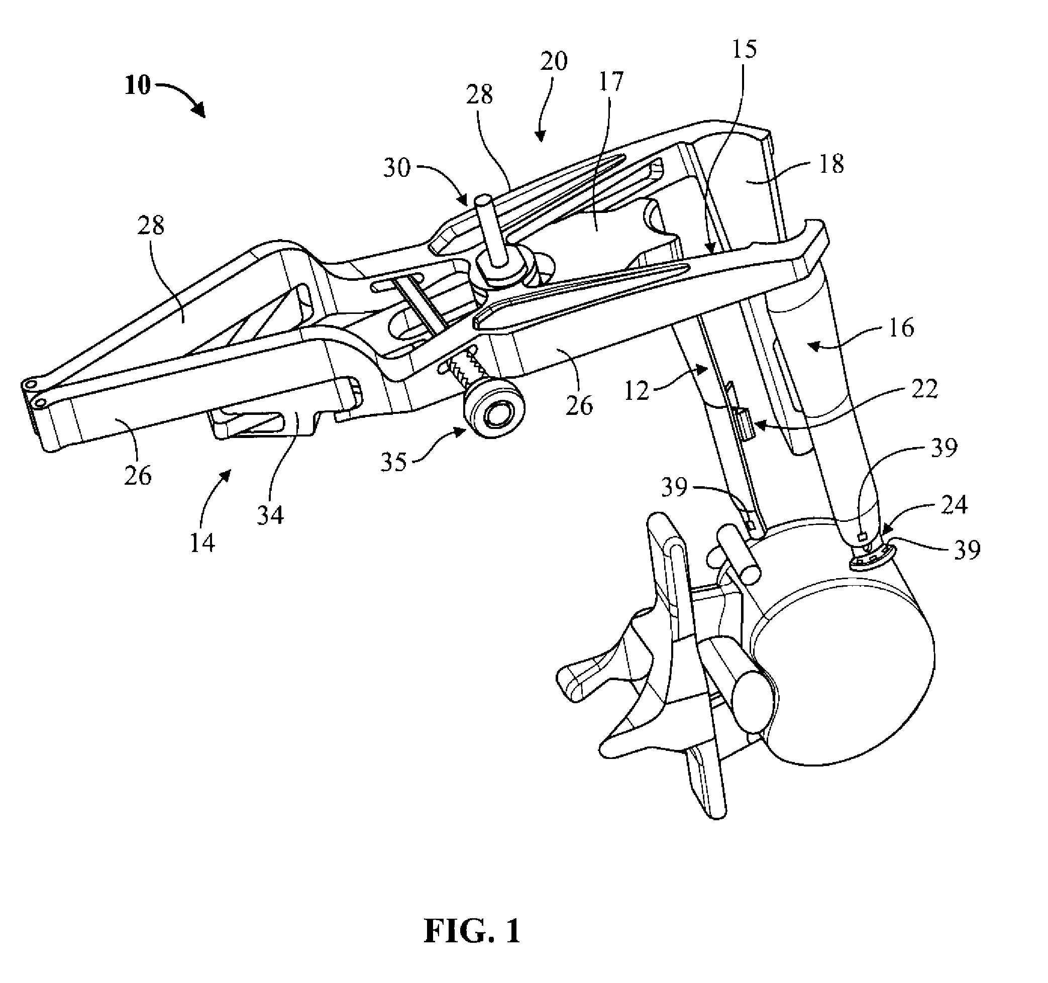
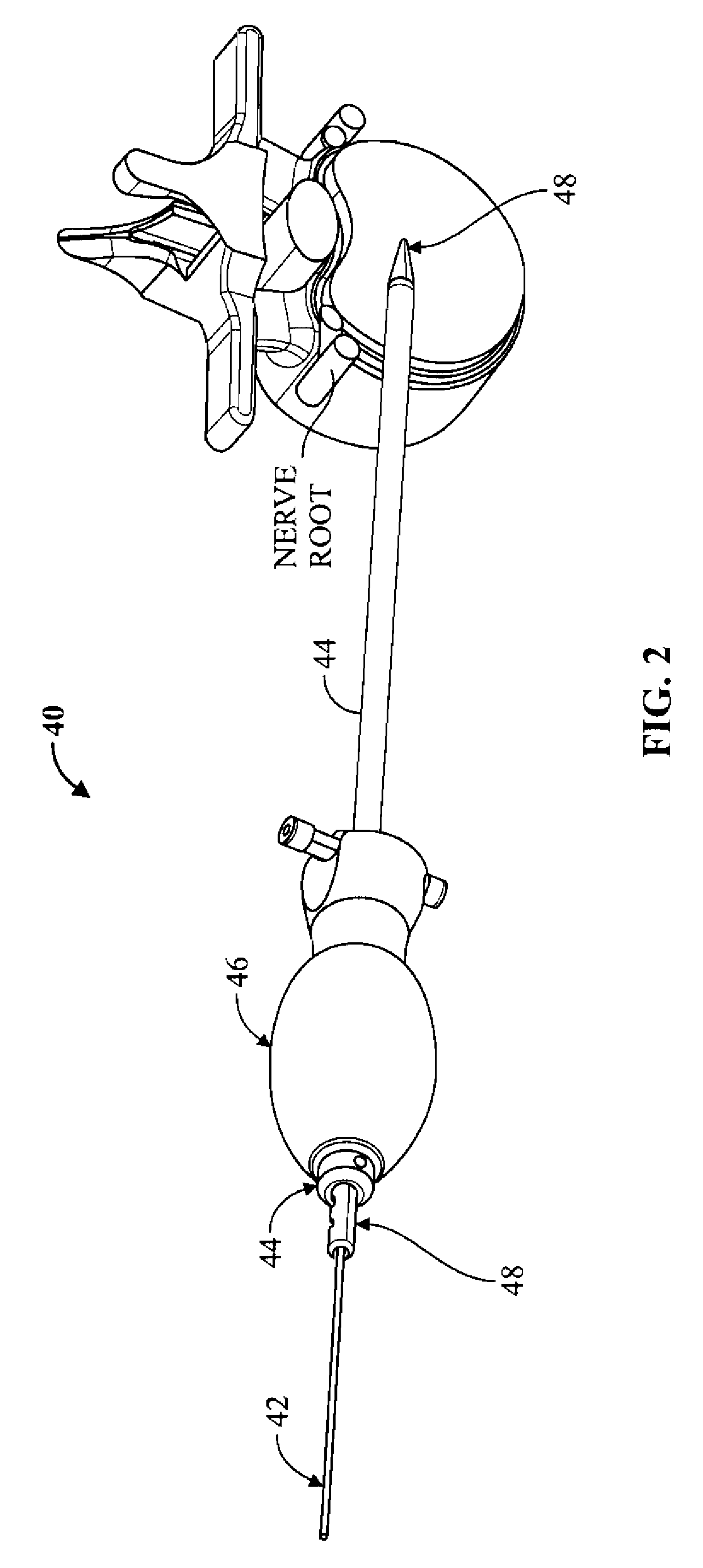
[0035]Illustrative embodiments of the invention are described below. In the interest of clarity, not all features of an actual implementation are described in this specification. It will of course be appreciated that in the development of any such actual embodiment, numerous implementation-specific decisions must be made to achieve the developers' specific goals, such as compliance with system-related and business-related constraints, which will vary from one implementation to another. Moreover, it will be appreciated that such a development effort might be complex and time-consuming, but would nevertheless be a routine undertaking for those of ordinary skill in the art having the benefit of this disclosure. It is furthermore to be readily understood that, although discussed below primarily within the context of spinal surgery, the surgical access system of the present invention may be employed in any number of anatomical settings to provide access to any number of different surgica...


PUM


Abstract

A surgical access system including a tissue distraction assembly and a tissue refraction assembly, both of which may be equipped with one or more electrodes for use in detecting the existence of (and optionally the distance and / or direction to) neural structures before, during, and after the establishment of an operative corridor to a surgical target site.

Description

CROSS-REFERENCES TO RELATED APPLICATIONS[0001]This application is a division of U.S. patent application Ser. No. 10 / 789,797, filed on Feb. 27, 2004, which claims priority to U.S. Provisional Patent Application Ser. No. 60 / 450,806, filed Feb. 27, 2003, the entire contents of which are hereby expressly incorporated by reference into this disclosure as if set forth fully herein.BACKGROUND OF THE INVENTION[0002]I. Field of the Invention[0003]The present invention relates generally to systems and methods for performing surgical procedures and, more particularly, for accessing a surgical target site in order to perform surgical procedures.[0004]II. Discussion of the Prior Art[0005]A noteworthy trend in the medical community is the move away from performing surgery via traditional “open” techniques in favor of minimally invasive or minimal access techniques. Open surgical techniques are generally undesirable in that they typically require large incisions and high amounts of tissue displace...

Claims


Application Information

Patent Timeline
Patent Type & Authority Applications(United States)
IPC IPC(8): A61B5/0488A61B1/32
CPCA61B17/02A61B1/32A61B5/04001A61B2017/0256A61B17/0206A61B17/0218A61B5/4836A61B5/24
Inventor MILES, PATRICKMARTINELLI, SCOTFINLEY, ERICGHARIB, JAMESFARQUHAR, ALLENKAULA, NORBERTBLEWETT, JEFFREYMEDEIROS, GORETTI
Owner NUVASIVE
Why Eureka
Try Eureka