Method and a device for imaging a visco-elastic medium

a viscoelastic medium and imaging method technology, applied in the field of methods and devices for imaging viscoelastic medium, can solve the problem of not providing any graduation in the rheology of lesions, and achieve the effect of tracking the variation in the size of a necrosis

Active Publication Date: 2010-07-01
SUPER SONIC IMAGINE
View PDF20 Cites 58 Cited by
  • Summary
  • Abstract
  • Description
  • Claims
  • Application Information

AI Technical Summary

Benefits of technology

[0033]Such a characteristic makes it possible to track variation in the visco-elastic properties of the medium as a function of time. In particular, this

Problems solved by technology

Finally, the known method serves only to detect the presence of liq

Method used


Image

  • Method and a device for imaging a visco-elastic medium
  • Method and a device for imaging a visco-elastic medium
  • Method and a device for imaging a visco-elastic medium

Examples


Embodiment Construction

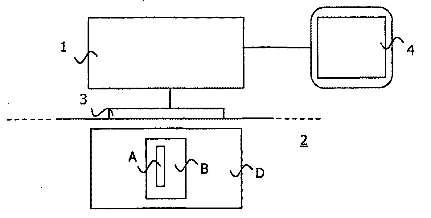
[0056]FIG. 1 is a diagrammatic representation of an imaging device 1 in accordance with the invention for imaging a visco-elastic medium 2. In an advantageous application, the medium2 is biological tissue, for example a human organ or portion of an organ, such as the breast.

[0057]The device 1 is connected to at least one ultrasound probe 3. Such a probe 3 may comprise a single element or a one-dimensional or a two-dimensional array of transducers. While the device of the invention is in use for observing the medium 2, the probe 3 is in contact with the medium 2.

[0058]The device 1 includes electronic means for controlling the emission of compression waves, e.g. ultrasound waves, by the probe 3.

[0059]The visco-elastic medium 2 diffuses such compression waves. In particular, ultrasound compression waves can propagate therein, thus enabling an echographic image to be made.

[0060]Advantageously, the device 1 is connected to a display module 4 enabling information extracted from the imagin...


PUM


Abstract

The invention relates to a method and a device for imaging a visco-elastic medium. The method comprises an excitation step during which an internal mechanical stress is generated in excitation zone and an imaging step of acquiring signals during the movements generated by the mechanical stress in the visco-elastic medium in response to the internal mechanical stress in an imaging zone that includes the excitation zone. According to the invention, the method further comprises a step of calculating a quantitative index associated with the rheological properties of the visco-elastic medium at at least one point of the imaging zone situated at a given depth outside the excitation zone. The quantitative index is representative of a comparison between signals acquired during the movements generated in response to the mechanical stress at at least one point of the excitation zone situated at the given depth, and signals acquired during the movements generated in response to the mechanical stress at at least the point of the imaging zone situated outside the excitation zone.

Description

BACKGROUND OF THE INVENTION[0001]The present invention relates to the general field of methods and devices for imaging a visco-elastic medium and including studying movements that exist in the visco-elastic medium. More particularly, the invention relates to methods comprising an excitation step during which an internal mechanical stress is generated in an excitation zone, followed by a step of imaging by acquiring signals during the movements generated in the visco-elastic medium in response to the internal mechanical stress in an imaging zone which includes the excitation zone.[0002]In a particularly advantageous application, the invention relates to imaging the visco-elastic properties of human organs. In this medical application, the invention serves in particular to detect the presence of a liquid zone in an organ such as the breast, the liver, the ovary, etc. and to characterize that zone.[0003]Echographic examination of the breast already makes it possible to deduce certain c...

Claims


Application Information

Patent Timeline
IPC IPC(8): A61B8/00
CPCA61B8/08A61B8/0825A61B8/0833A61B8/485A61N7/00G01S7/52036G01S7/52042G01S15/8961G01S15/8977A61B5/0051
Inventor BERCOFF, JÉRÉMYSAVERY, DAVIDCOHEN-BACRIE, CLAUDESOUQUET, JACQUES
Owner SUPER SONIC IMAGINE
Why Eureka
Try Eureka