
Magnetic resonance method and apparatus using dual echoes for data acquisition

a magnetic resonance and data acquisition technology, applied in the field of magnetic resonance imaging methods and systems, can solve the problems of insufficient temporal resolution, inefficient acquisition scheme, and particularly problematic, and achieve the effect of improving the overall image quality

Active Publication Date: 2010-10-21
UK RES & INNOVATION LTD +1
View PDF21 Cites 10 Cited by
  • Summary
  • Abstract
  • Description
  • Claims
  • Application Information

AI Technical Summary

Benefits of technology

[0012]It is an object of the present invention to provide a magnetic resonance imaging method and apparatus that allows images of a region of a subject to be obtained with an improved overall image quality, despite at least a portion of the imaged region being susceptible to inhomogeneities of the basic magnetic field of the magnetic resonance imaging apparatus.
[0013]It is a further object of the present invention to provide such a method and apparatus that allows improved brain images to be obtained in fMRI.

Problems solved by technology

For most fMRI applications, however, this acquisition scheme is suboptimal for a number of reasons.
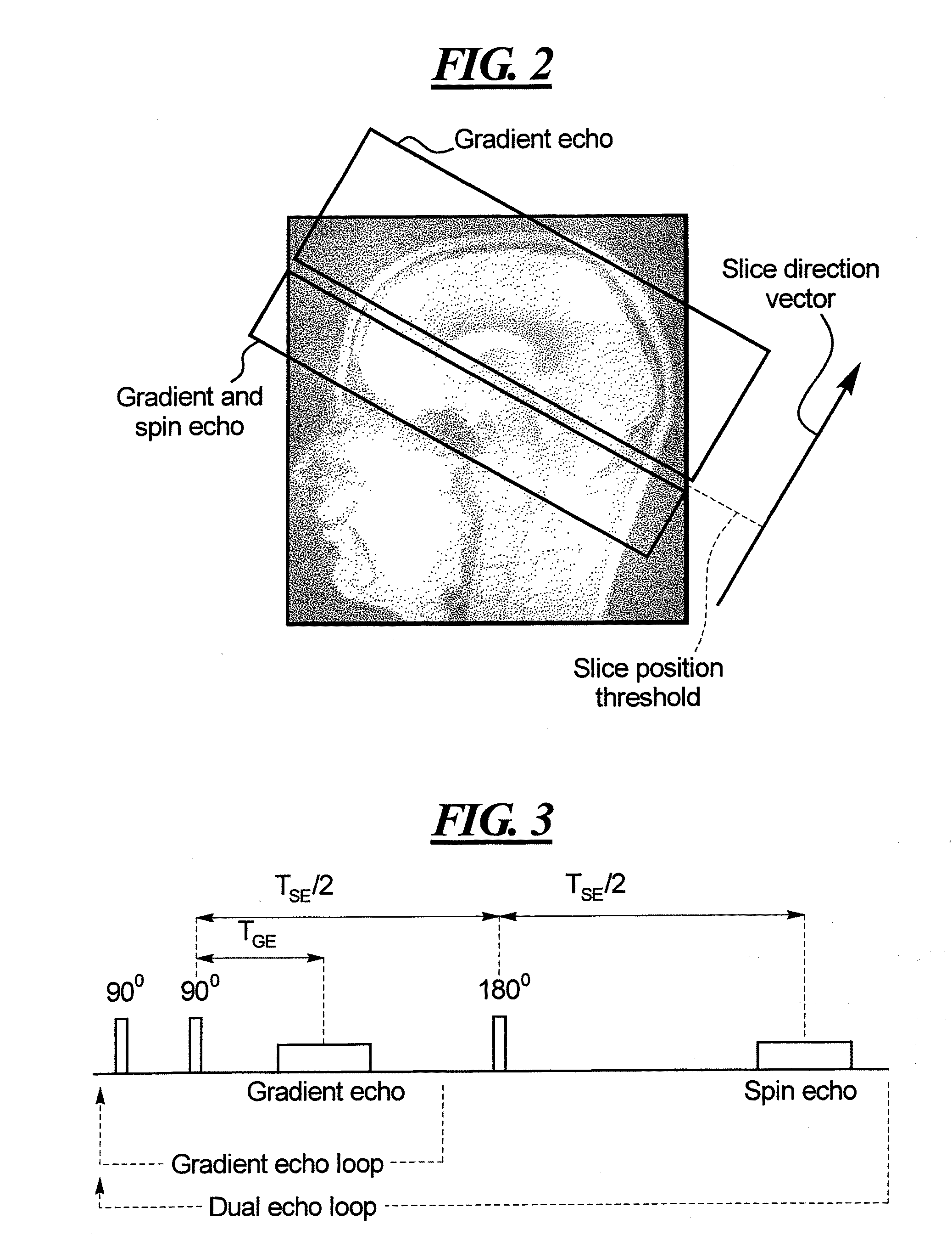
The main drawbacks of using the single shot dual echo EPI sequence for fMRI are:(1) More than a 100% increase in the minimum repetition time (TR), because two images are acquired per slice.(2) The resulting temporal resolution is often insufficient for event-related fMRI designs.(3) The acquisition scheme is inefficient as signal losses due to magnetic field inhomogeneity typically only occur in the lower third of the brain.(4) The resulting time normalized SNR is suboptimal (meaning SNR normalized by the square root of the total acquisition time).(5) The 180° refocusing pulses lead to a substantial increase in the SAR (specific absorption rate).
This may be particularly problematic at high and ultrahigh field strength.

Method used


Image

  • Magnetic resonance method and apparatus using dual echoes for data acquisition
  • Magnetic resonance method and apparatus using dual echoes for data acquisition
  • Magnetic resonance method and apparatus using dual echoes for data acquisition

Examples


Embodiment Construction

[0023]FIG. 6 is a block diagram schematically illustrating the basic components of a magnetic resonance imaging system that is suitable for implementing the method in accordance with the present invention. The basic structure of the components is known, but either or both of the system computer 20 and the sequence control 18 is / are appropriately programmed with a control protocol for operating the system in accordance with the present invention.

[0024]A basic field magnet 1 generates a temporally constant, strong magnetic field for polarization or alignment of the nuclear spins in the examination region of a subject (such as, for example, a portion of a human body to be examined). The high homogeneity of the basic magnetic field that is required for the nuclear magnetic resonance measurement is defined in a spherical measurement volume M into which the portions of the human body to be examined are introduced. Components known as shim plates (not shown) made from ferromagnetic materia...


PUM


Abstract

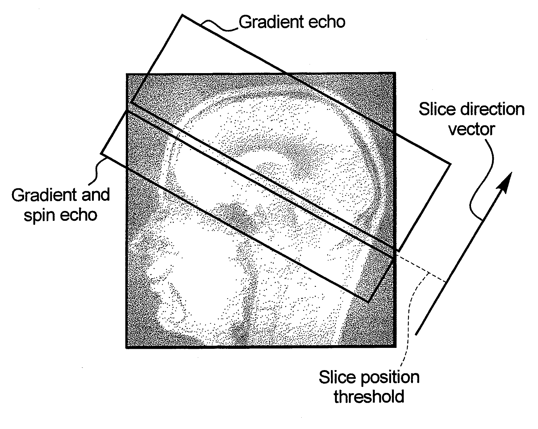
In a method and system for acquiring magnetic resonance image data from a subject, using a data acquisition unit in which a basic magnetic field is generated, a division is designated in a planar section through the subject that divides the planar section into a first section and a second section, with the homogeneity of the basic magnetic field being better in said first section than in said second section. An echo planar imaging sequence is implemented to acquire the magnetic resonance imaging data, with magnetic resonance data being acquired from the first section only from gradient echo signals in the echo planar imaging sequence, and magnetic resonance data from said the second section being acquired from both gradient echo signals and spin echo signals in the echo planar imaging sequence. The method and apparatus are particularly suited for acquiring functional magnetic resonance imaging (fMRI) data from the brain of a subject, in which the first section is an upper portion of the brain and the second section is a lower portion of the brain.

Description

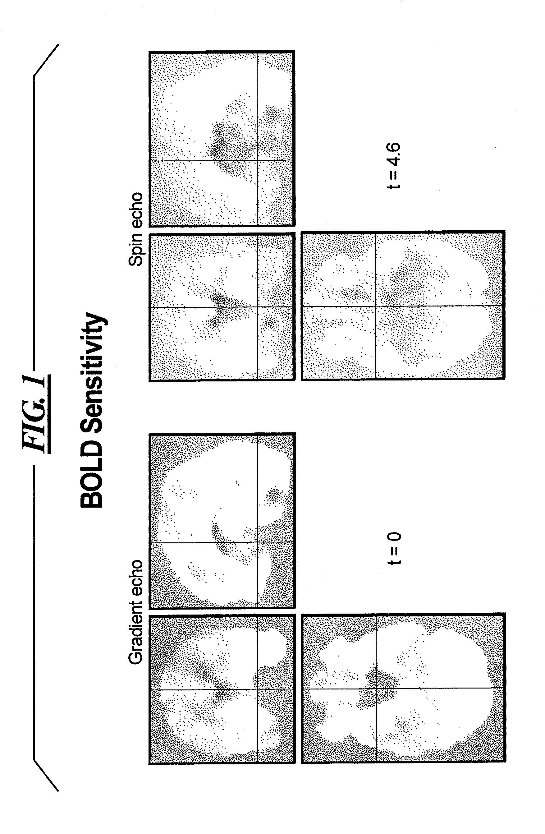
BACKGROUND OF THE INVENTION[0001]1. Field of the Invention[0002]The present invention relates to methods and systems for magnetic resonance imaging, and in particular to methods and systems wherein magnetic resonance image data are acquired using an echo planar imaging (EPI) pulse sequence.[0003]2. Description of the Prior Art[0004]Most functional magnetic resonance imaging (fMRI) studies are based on the gradient echo EPI (GE-EPI). In general, this technique is known to be more sensitive to changes in the known BOLD (Blood, Oxygen Level Dependent) contrast than spin echo EPI (SE-EPI). On the other hand, SE-EPI is considerably more robust against signal losses or voids caused by magnetic field inhomogeneities, which typically occur in brain regions near air-tissue interfaces, such as the orbito-frontal cortex or the inferior temporal lobes, as described in Bandettini et al., “Spin-Echo and Gradient-Echo EPI of Human Brain Activation Using Bold Contrast—A Comparative-Study at 1.5 T” ...

Claims


Application Information

Patent Timeline
IPC IPC(8): A61B5/055
CPCG01R33/4806G01R33/56563G01R33/5618A61B5/055A61B2018/00446
Inventor PORTER, DAVID ANDREWSCHWARZBAUER, CHRISTIAN
Owner UK RES & INNOVATION LTD
Learn More