Genome-wide analysis of palindrome formation and DNA methylation

a gene and methylation technology, applied in the field of gene-wide analysis of palindrome formation and dna methylation, can solve the problems of limited examples, dna preparation, and inability to detect dsb repair,

Inactive Publication Date: 2010-10-28
FRED HUTCHINSON CANCER RES CENT
View PDF12 Cites 28 Cited by
  • Summary
  • Abstract
  • Description
  • Claims
  • Application Information

AI Technical Summary

Problems solved by technology

Moreover in mice deficient in both p.
This indicates that improper DSB repair also could trigger the BFB cycle for further chromosome aberrations.
However, our molecular analysis of the structure of amplified loci in cancer cells has been limited by the fact that the duplication covers very large regions of the chromosome.
However, these examples are limited in number and fail to reveal the full scope of dynamic changes in methylation status.
These methods can be disadvantageous because each method is dependent on the presence and optimal spacing of methylation sensitive restriction enzyme recognition sites and variable methylation patterns with similar densities can cause differential signals.

Method used


Image

  • Genome-wide analysis of palindrome formation and DNA methylation
  • Genome-wide analysis of palindrome formation and DNA methylation
  • Genome-wide analysis of palindrome formation and DNA methylation

Examples


example 1

[0063]The following example describes a process for genome-wide assessment of palindrome formation.

Methods

Cell Lines and Cancer Tissues

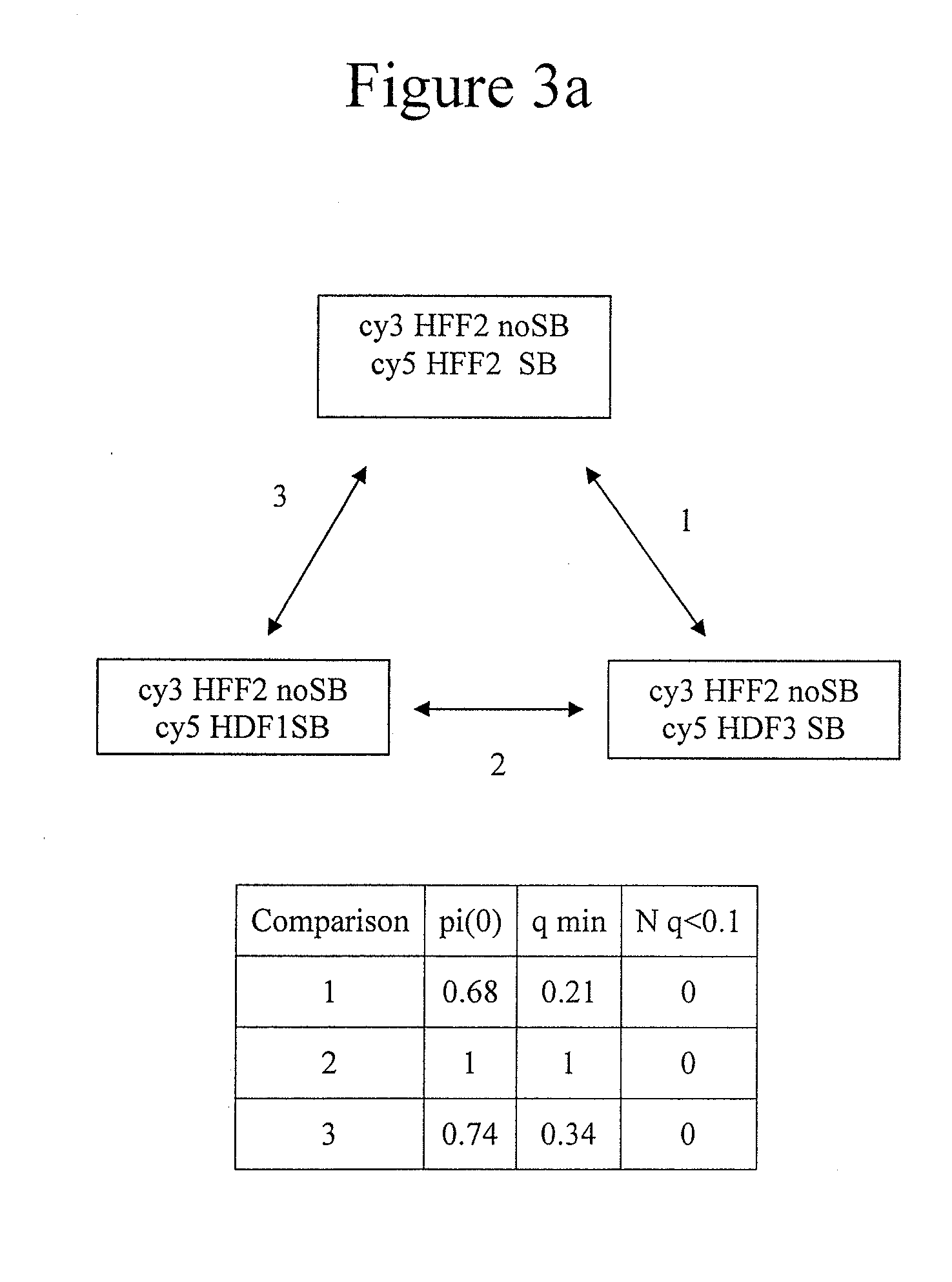
[0064]D79IR-8 and D79IR-8-Sce 2 cells were previously described (Tanaka et al., Proc. Natl. Acad. Sci. USA 99:8772-8777 (2002)). Colo320DM and RD were obtained from American Type Culture Collection. MCF7 and AG 1113215 were from the University of Washington. Skin biopsy derived fibroblasts HDF1 and HDF3 were obtained from the University of Washington and human foreskin fibroblasts HFF2 from the Fred Hutchinson Cancer Research Center (FHCRC) as anonymous cell lines. DNA samples stripped of identifying information from five primary medulloblastomas were provided by the Fred Hutchinson Cancer Research Center. All samples were obtained after Fred Hutchinson Cancer Research Center Institutional Review Board review and approval for use of anonymous human DNA samples and human cell lines.

Linkers and Oligonucleotides

[0065]Oligonucleotides were synthesized by...

example 2

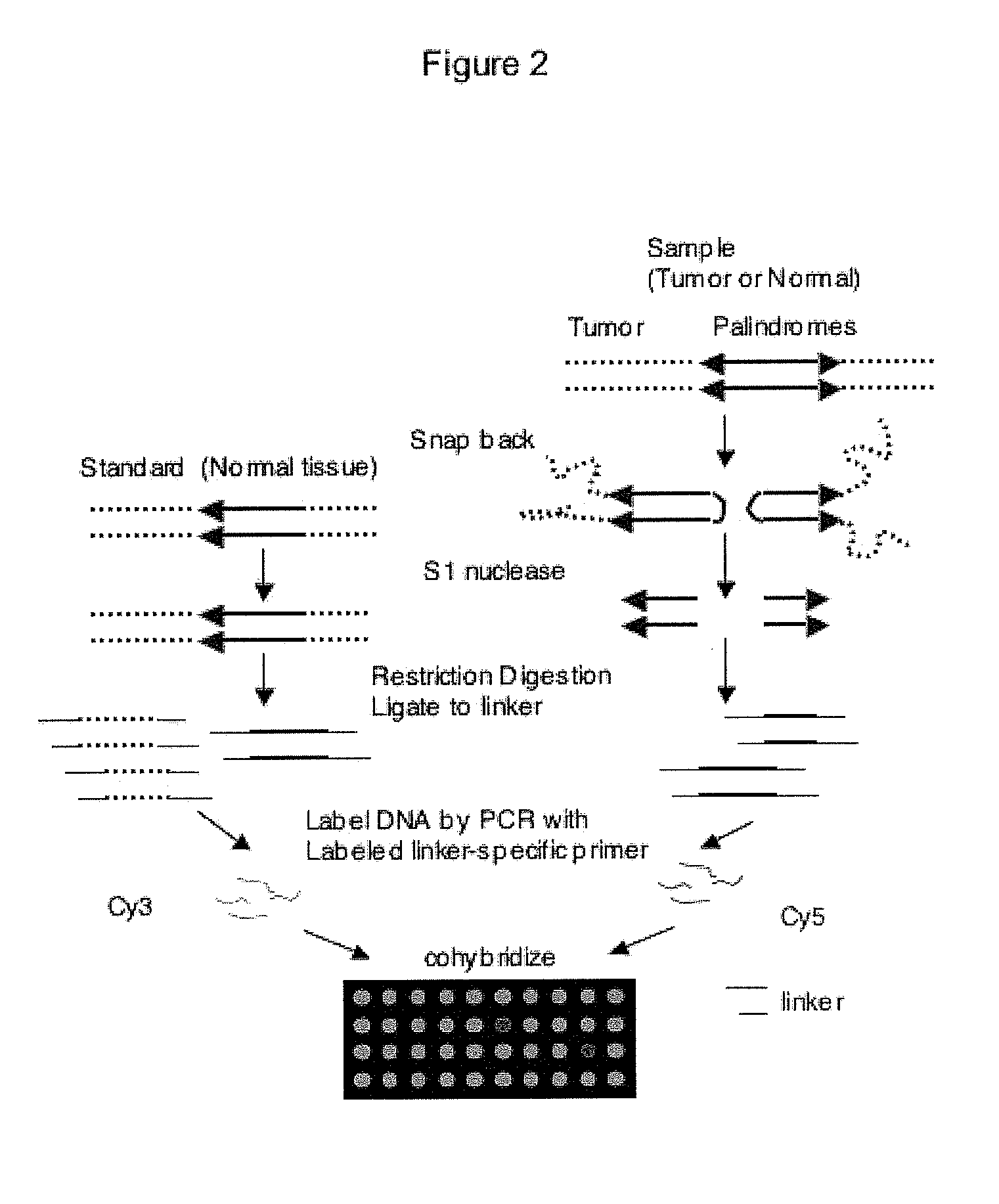
[0086]The following example demonstrates the use of ligation-mediated PCR to isolate a DNA fragment enriched in unmethylated CpG islands in a mammalian cell. A schematic of the process is provided as FIG. 8A.

[0087]Briefly, mouse genomic DNA was digested with a methylation sensitive restriction enzyme (for example, HpaII). The MspI linkers used above in Example 1 were used to ligate the HpaII fragments. The ligated DNA was amplified by PCR using the MspI primer from Example 1 (SEQ ID NO: 6). The method resulted in the specific amplification of HpaII digested genomic DNA of less than 500 base pairs (FIG. 8B). Random cloning and sequencing of the PCR products revealed that more than 50% of clones were at the CpG islands as defined using stringent criteria. (Takai and Jones, Proc. Natl. Acad. Sci. USA 99:3740-3745 (2002); incorporated herein by reference). In contrast, amplification of DNA digested with methylation-resistant isoschizomer MspI gave no clones near CpG islands.

TABLE 1Resul...

example 3

[0089]The following example describes methods used to identify palindromes and methylated DNA.

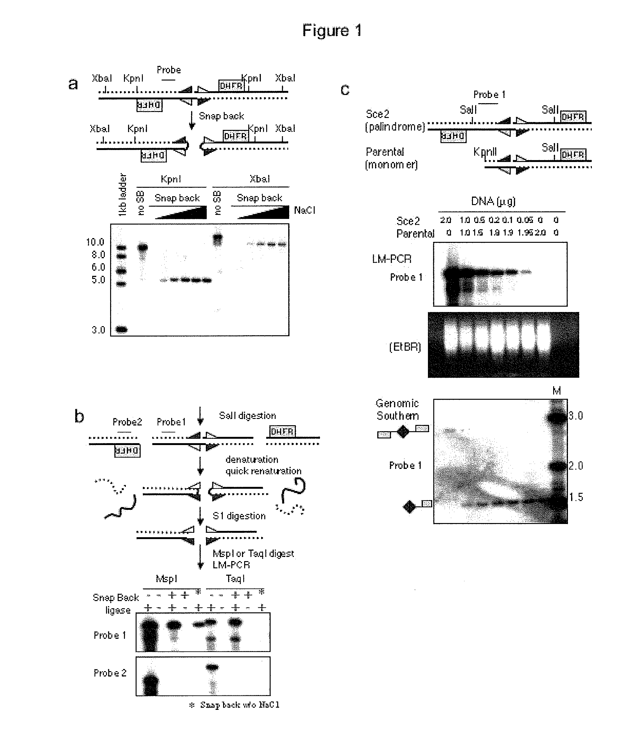
[0090]Above is described a method to obtain a genome-wide analysis of palindrome formation (GAPF) based on the efficient intrastrand base pairing in large palindromic sequences (Tanaka et al., Nat. Genet. 37:320-327 (2005)). Palindromic sequences can rapidly anneal intramolecularly to form ‘snap-back’ DNA under conditions that do not favor intermolecular annealing. This snap-back property was used to enrich for palindromic sequences in total genomic DNA by denaturing the DNA at 100° C. in the presence of 100 mM NaCl, rapidly renaturing it by snap cooling, and then digesting the mixture with a single-strand specific nuclease. Snap-back DNA formed from palindromes was double-stranded and resistant to the single-strand specific nuclease, whereas the remainder of genomic DNA was single-stranded and thus was sensitive to digestion (FIG. 9). Using this assay, de novo palindromes were shown to for...


PUM

PropertyMeasurementUnit
melting temperatureaaaaaaaaaa
temperaturesaaaaaaaaaa

Abstract

The present disclosure provides methods for detecting the genome-wide presence of methylated DNA and palindrome formation. The present disclosure also provides methods for specific enrichment of methylated DNA or DNA having a DNA palindrome. These methods have demonstrated that somatic palindromes and methylated DNA occur frequently and are widespread in human cancers. Individual tumor types have a characteristic non-random distribution of palindromes in their genome and a small subset of the palindromic loci are associate with gene amplification. The disclosed method can be used to define the plurality of genomic DNA palindromes and regions having methylated DNA associated with various tumor types and can provide methods for the classification of tumors, and the diagnosis, early detection of cancer as well as the monitoring of disease recurrence and assessment of residual disease.

Description

CROSS-REFERENCES TO RELATED APPLICATIONS[0001]The present application is a continuation-in-part of U.S. patent application Ser. No. 11 / 142,091, which claims priority to U.S. Provisional Patent Application No. 60 / 575,331, filed May 28, 2004, the entire disclosures of which are incorporated by reference herein.STATEMENT AS TO RIGHTS TO INVENTIONS MADE UNDER FEDERALLY SPONSORED RESEARCH OR DEVELOPMENT[0002]This invention was made with government support under Grant Nos. R01AR 045113, R01GM 26210, K12 HD43376 and 2T32CA009351 awarded by the National Institutes of Health. The Government has certain rights in the invention.BACKGROUND[0003]Cancer is a disease of impaired genetic integrity. In most cases disturbed genetic integrity is observed at the chromosome level and include a configuration called anaphase bridges, which are most likely derived from dicentric or ring chromosomes segregating into two different daughter cells in the process of the breakage-fusion-bridge (BFB) cycle. The B...

Claims


Application Information

Patent Timeline
Patent Type & Authority Applications(United States)
IPC IPC(8): C12Q1/68
CPCC12N15/1093C12Q1/6827C12Q1/6886C12Q2600/16C12Q2600/154C12Q2600/112C12Q2537/164C12Q2527/107C12Q2521/307
Inventor TAPSCOTT, STEPHEN J.TANAKA, HISASHIYAO, MENG-CHAODIEDE, SCOTT J.
Owner FRED HUTCHINSON CANCER RES CENT
Why Eureka
Try Eureka