Method for the improvement of mesh implant biocompatibility

a mesh implant and biocompatibility technology, applied in the field of surgery, can solve the problems of chronic pain and adhesion formation, seroma formation, and impaired abdominal wall function, and achieve the effect of reducing the risk of adhesion formation

Active Publication Date: 2010-11-18
BAXTER INT INC +1
View PDF102 Cites 100 Cited by
  • Summary
  • Abstract
  • Description
  • Claims
  • Application Information

AI Technical Summary

Benefits of technology

[0007]The invention also relates to a method of reducing the risk of adhesion formation after surgery to a mesh implant in a subject comprising attaching said mesh implant to a tissue of the subject and covering said mesh impla

Problems solved by technology

Complications found with IPOM technique are seroma formation, impaired functionality of the abdominal wall, chronic pain and adhesion formation.
These adhesions are potentially resulting in major complications like bowel

Method used


Image

  • Method for the improvement of mesh implant biocompatibility
  • Method for the improvement of mesh implant biocompatibility
  • Method for the improvement of mesh implant biocompatibility

Examples


example 1

Use of an Anionic Polysaccharide

[0040]Male Sprague Dawley rats, weighing 400-500 g are used.

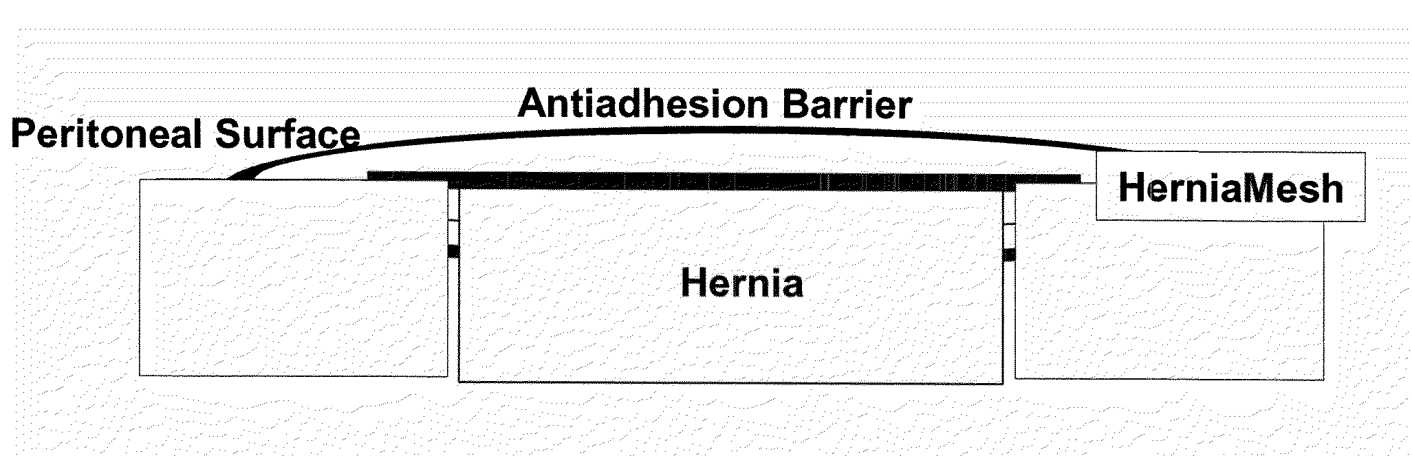
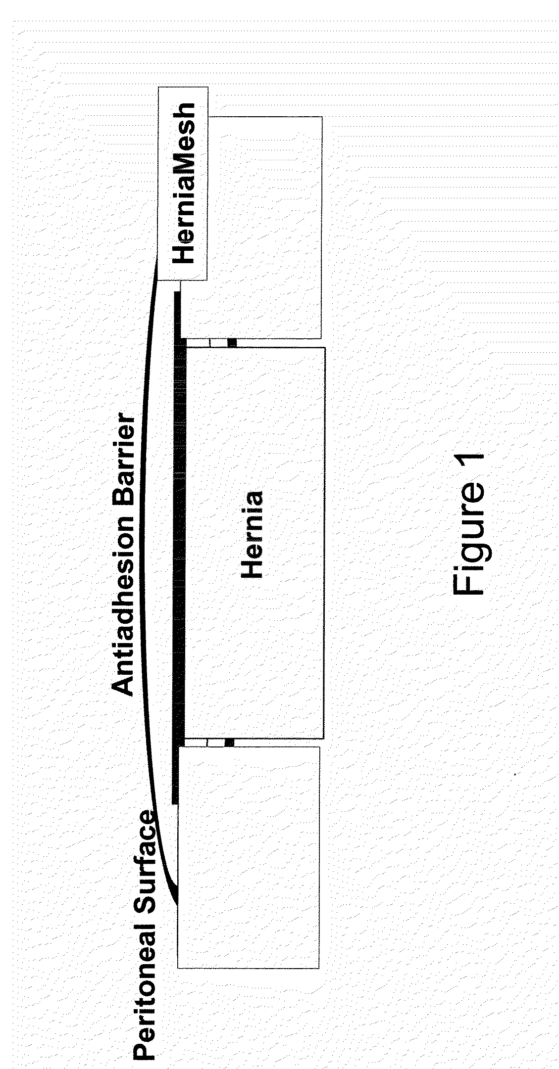
[0041]A macroporous, middleweight polypropylene mesh is used in the trial. This mesh should not be used intraabdominally without antiadhesive barriers. Polypropylene mesh only serves as control group.

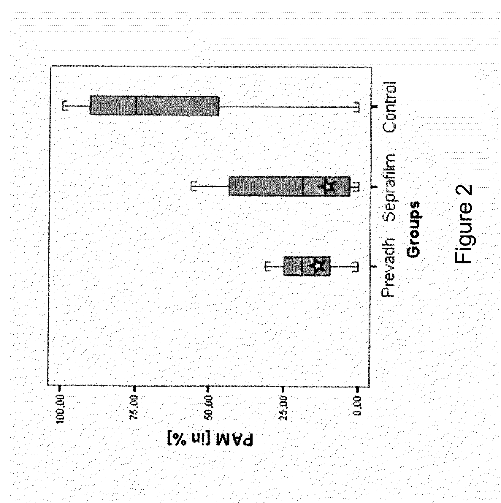
[0042]The antiadhesive barrier tested in combination with VM consists of anionic polysaccharides (Seprafilm®). The choice of the antiadhesive barrier is based on experimental and clinical studies showing an antiadhesive effect and good biocompatibility of the materials as well as on their availability to be purchased separately.

[0043]Observation time is 30 days to assure completed wound healing in order to be able to examine tissue integration and long-term complications like seroma and adhesion formation. Surgery is performed under sterile conditions.

[0044]Rats are anaesthetized with an ip injection of 110 mg / kg BW of Ketavet® (Ketamine-hydrochloride 100 mg / ml, Pharmacia, Germany) and 12 mg / kg B...

example 2

Use of a Collagen Membrane

[0049]The setup is the one as described in Example 1, but the antiadhesive barrier tested in combination with VM consists of atelocollagen type 1 (Prevadh®).

example 3

Use of Amniotic Membrane

[0050]Besides the various commercially available antiadhesive barriers which are specifically designed for the combination with synthetic hernia meshes, preliminary findings indicate a potential of sealed AM for this indication. Due to the biomechanical characteristics of AM, the technical difficulties concerning application to the mesh and fixation are similar to the use of other organic barriers and coatings.

[0051]The efficacy of FS to securely fit and fix AM to a polypropylene mesh in our previously published IPOM model in rats (n=14). The AM is checked for vitality prior to implantation and pre-cut to squares of 2×2 cm. These samples are attached to VM by means of 0.2 ml Artiss (4 I.U.) each. The mesh is additionally fixated with 4 corner sutures.

[0052]Antiadhesive properties of AM are shown and AM is securely attached to the mesh after an observation period of 14 days. FS is fully degraded and neovascularisation is detected histologically. Our preliminar...


PUM


Abstract

The present invention provides a method of fixating a mesh implant to a tissue of a subject comprising attaching said mesh implant to said tissue, covering said mesh implant by an antiadhesive barrier, wherein said antiadhesive barrier is attached to said mesh implant by a biocompatible adhesive.

Description

FIELD OF THE INVENTION[0001]The present invention relates to the field of surgery, in particular the integration of artificial mesh implants.BACKGROUND OF THE INVENTION[0002]The placement of synthetic meshes into the body of a patient has become routine in the field of surgical practice. The most common use for mesh implants is the placement into the abdominal cavity for the repair of ventral and incisional hernias. The meshes are either placed extraperitoneally in onlay or sublay technique or intraperitoneally in intraperitoneal onlay mesh technique (IPOM). Inlay technique also played a role in the past. IPOM technique is indicated in multiple defect hernias (also known as Swiss cheese hernias) as well as for the treatment of large abdominal wall defects with loss of domain in obese patients. The implants are usually fixed by sutures, tacks or anchors. Complications found with IPOM technique are seroma formation, impaired functionality of the abdominal wall, chronic pain and adhesi...

Claims


Application Information

Patent Timeline
IPC IPC(8): A61B17/03
CPCA61B2019/4884A61F2/0063A61L31/041A61F2002/009A61F2/0077A61B2090/0816
Inventor PETTER-PUCHNER, ALEXANDERREDL, HEINZ
Owner BAXTER INT INC
Why Eureka
Try Eureka