
System and method for storing and activating an in vivo imaging capsule

a technology of imaging capsules and storage methods, applied in the field of in vivo devices and methods, can solve the problems of reducing unable to retrieve or activate the capsules, and affecting the quality of images captured by the imaging capsules

Active Publication Date: 2010-12-02
GIVEN IMAGING LTD
View PDF51 Cites 16 Cited by
  • Summary
  • Abstract
  • Description
  • Claims
  • Application Information

AI Technical Summary

Benefits of technology

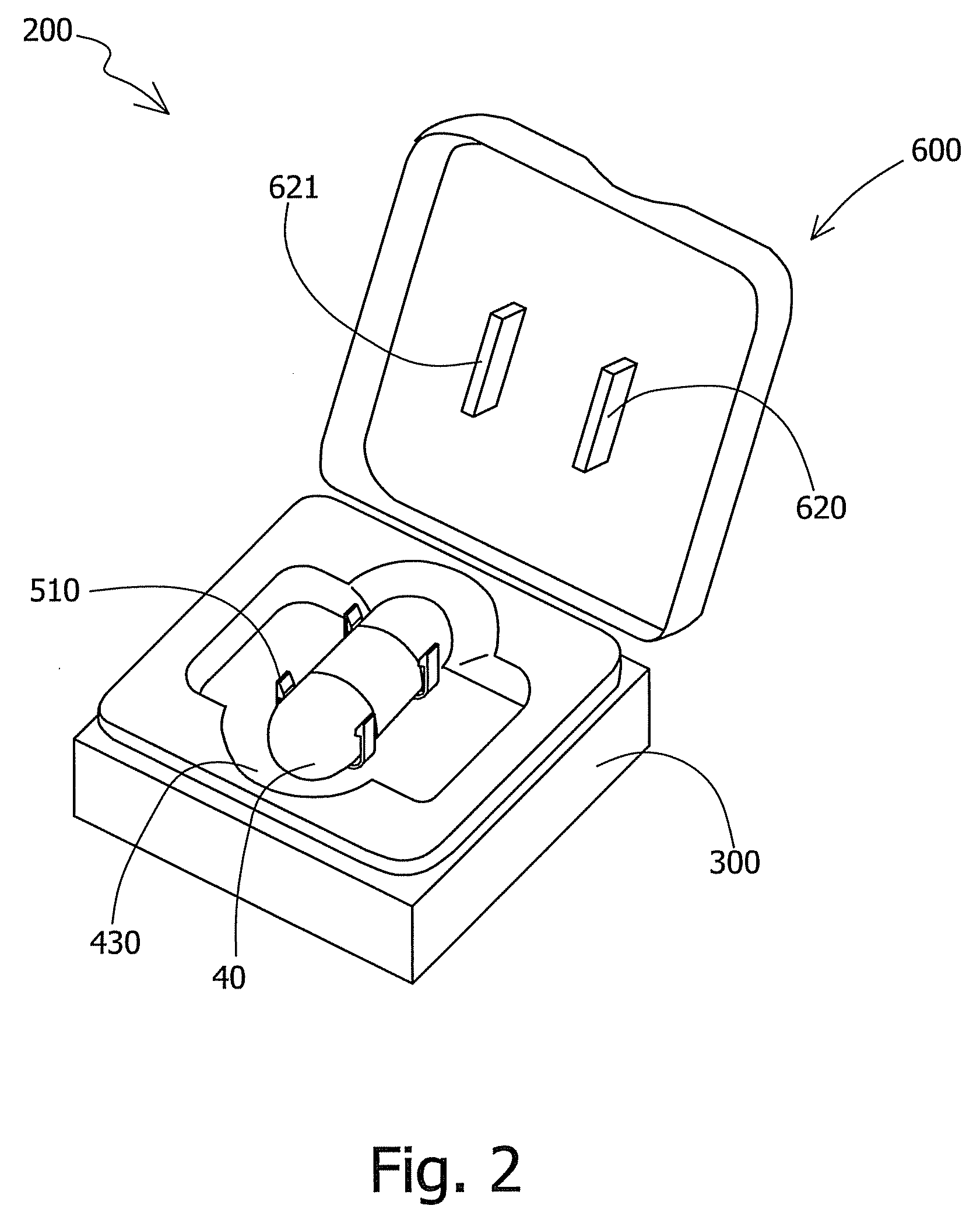
[0011]In one embodiment, the capsule holding unit may comprise one or more capsule gripping units. Each capsule gripping unit may comprise two protruding elements, positioned for example substantially symmetrically across from each other on opposite sides of the recessed space of the storage unit. The capsule gripping units may be positioned substantially one centimeter apart from each other. In other embodiments, the capsule gripping units may be positioned differently, for example closed together or further apart.
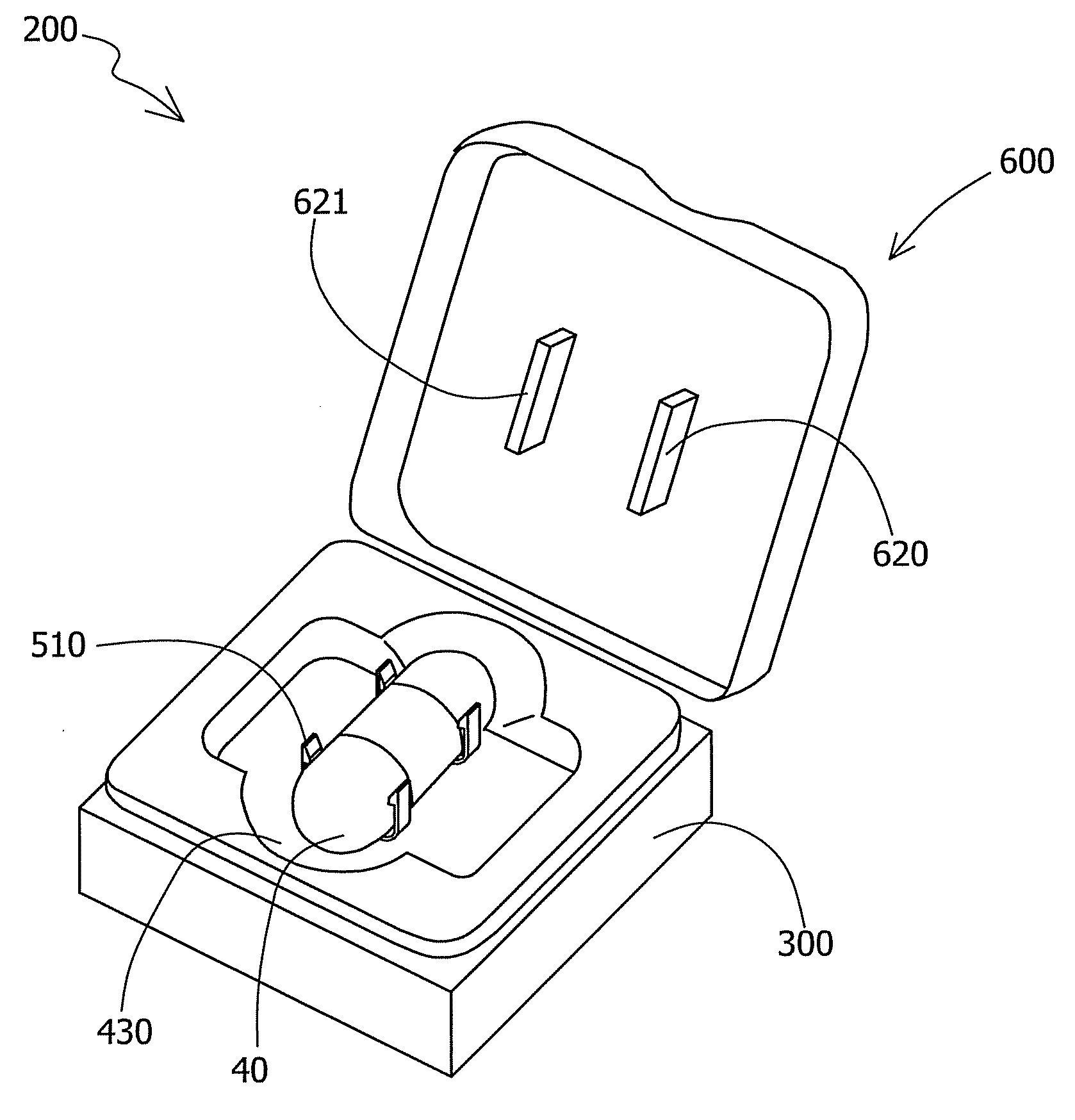
[0012]A storage device is provided according to one embodiment for storing a swallowable capsule. The storage device may comprise, in one embodiment, a container base with a recessed space for placing the swallowable capsule, a capsule holding unit which may be positioned for example substantially within in the container base, and a lid for covering the container base. The lid may comprise a magnet, which may be positioned to activate the swallowable capsule when the lid is open, and to deactivate the swallowable capsule when the lid is closed. The capsule holding unit may comprise for example two protruding elements, which may be positioned to removably hold the swallowable capsule around its housing. The lid may comprise a fastening element to press on the two protruding elements when the lid is closed, causing the swallowable capsule to be held in the capsule holding unit. The storage device may further comprise a liner, which may be assembled into the container base.

Problems solved by technology

In some cases, it is not possible to retrieve or to activate the capsule after it has been swallowed.
However, an imaging capsule should not be activated accidentally or involuntarily, for example during shipping and handling of the capsule in its package, since such activation may deplete the capsule's battery and cause the imaging procedure to fail.
The optical dome may be sensitive to scratches or deformation, which may occur during transportation, shipping and / or handling of the capsule in its package.
Deformation or dirt may obscure the imaging field of view, and / or may cause imaging artifacts which reduce the quality of the images captured by the imaging capsule.
Images with artifacts or with a reduced field of view may reduce the detectability of pathologies or abnormalities by the specialist reviewing the image stream.
A large dome may be more prone to damaging or accidental scratching, breaking or impairment.
Storing devices for example such as disclosed in U.S. Pat. No. 7,295,226 to Meron et al may be primarily designed, for example, for holding and protecting an imaging capsule with a single optical head, and may be less desirable for an imaging capsule with more than one optical head.
However, the act of removing an imaging capsule with for example two optical domes located at opposite ends of the capsule from such a storage device may cause the user to touch one of the optical domes which is not inserted into the cup.
The same dome may also be damaged during the shipping of the capsule in its package.

Method used


Image

  • System and method for storing and activating an in vivo imaging capsule
  • System and method for storing and activating an in vivo imaging capsule
  • System and method for storing and activating an in vivo imaging capsule

Examples


Embodiment Construction

[0027]In the following detailed description, numerous specific details are set forth in order to provide a thorough understanding of the present invention. However, it will be understood by those skilled in the art that the present invention may be practiced without these specific details. In other instances, well-known methods, procedures, and components have not been described in detail so as not to obscure the present invention.

[0028]Some embodiments of the present invention are directed to storing and activating an in-vivo device that may be inserted (e.g., by swallowing) into a body lumen, e.g., the gastro-intestinal (GI) tract, for example, from outside the body. Some embodiments are directed to a typically one time use or partially single use detection and / or analysis device. Some embodiments are directed to storing a typically swallowable in-vivo device that may passively or actively progress through a body lumen, e.g., the gastro-intestinal (GI) tract, for example, pushed a...


PUM


Abstract

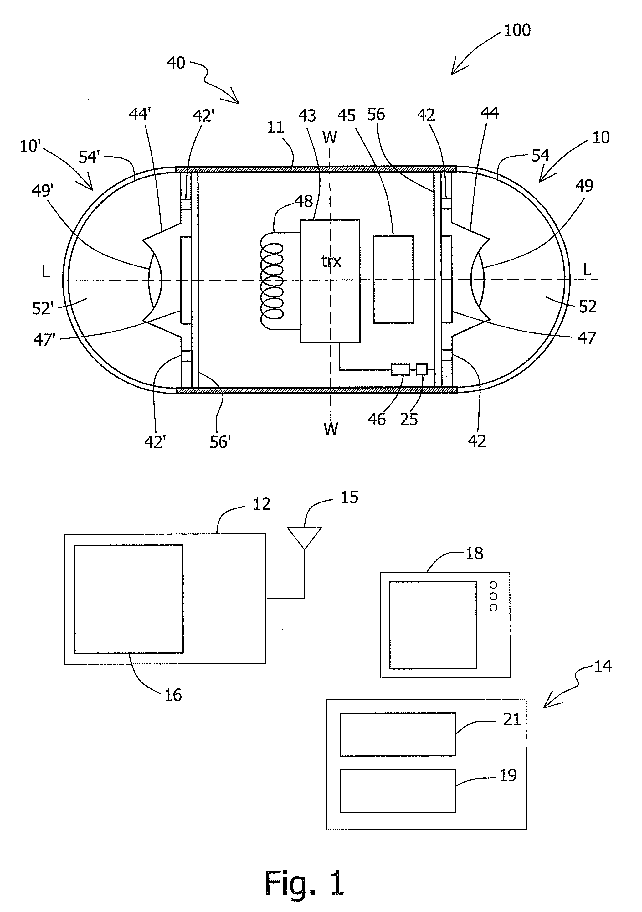
A storage unit is provided for removably storing an in vivo imaging capsule. The capsule may have a housing and an optical dome. The storage unit may include a recessed space formed for accepting the capsule. A magnet for magnetically activating and deactivating the capsule may be provided in the storage unit, for example in the lid. The storage unit may also include a capsule holding unit comprising at least two protruding elements to removably hold the capsule in the storage unit, wherein the protruding elements are positioned to grip the capsule housing.

Description

FIELD OF INVENTION[0001]The present invention relates to an in-vivo device and method such as for imaging an in-vivo lumen. More specifically, the present invention relates to a method and apparatus for storing and activating an in-vivo imaging capsule.BACKGROUND OF THE INVENTION[0002]Known devices may be helpful in in-vivo sensing, such as imaging or pH sensing. Autonomous in-vivo sensing devices, such as swallowable or ingestible capsules or other devices, may move through a body lumen, sensing as they move along. An autonomous in-vivo sensing device such as an imaging device may include, for example, an imager for obtaining images from inside a body cavity or lumen, such as the gastrointestinal (GI) tract while the in-vivo imaging device passes through the GI lumen. The imager may, for example, be associated with an optical system, and optionally a transceiver and an antenna. Some of these devices use a wireless connection to transmit image data. Other devices, systems and method...

Claims


Application Information

Patent Timeline
Patent Type & Authority Applications(United States)
IPC IPC(8): B65D85/42A61B1/06
CPCA61B1/041A61B1/00144
Inventor GILAD, ZVIKAROYTENBURD, VITALY
Owner GIVEN IMAGING LTD
Learn More