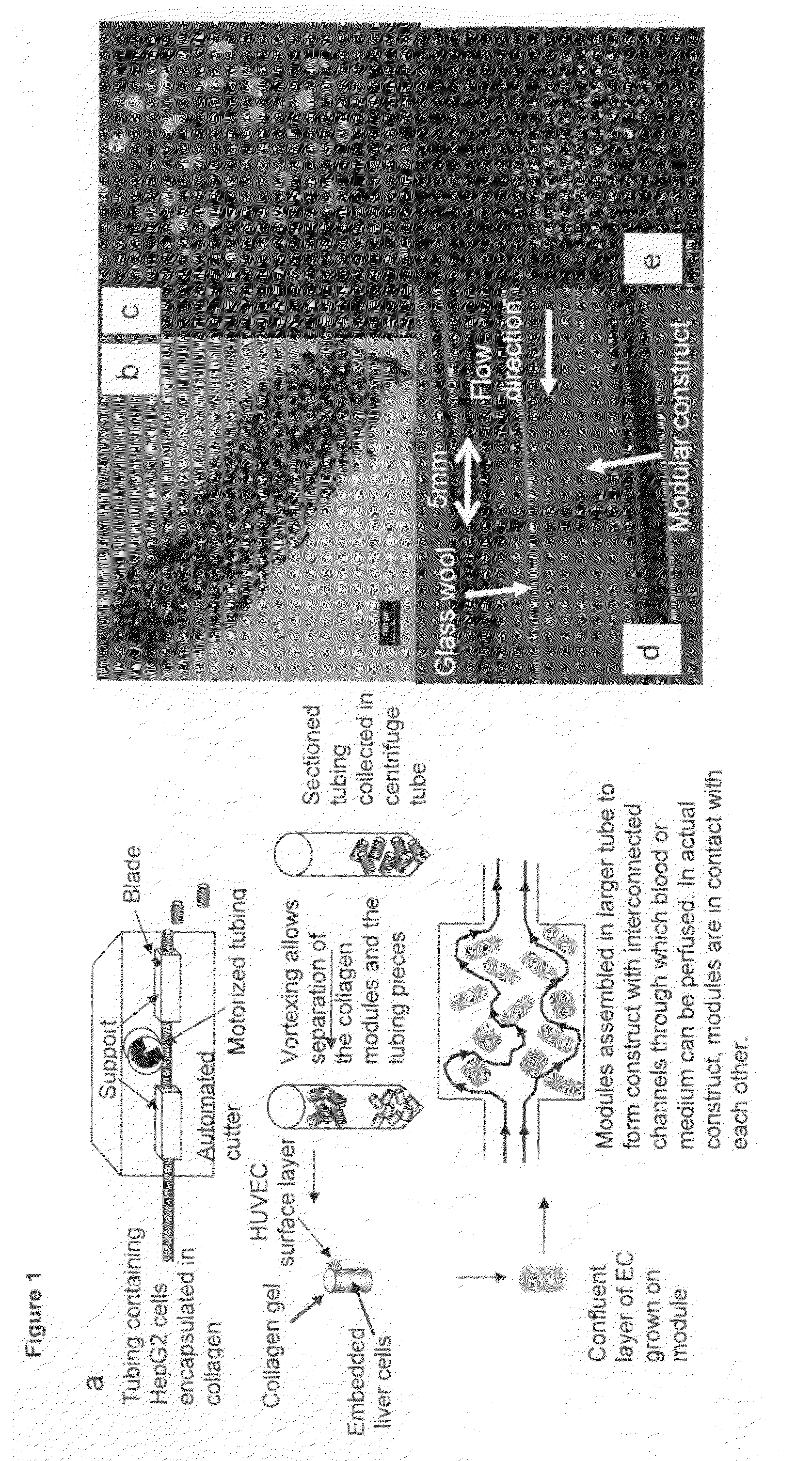
Modular assembly of tissue engineered constructs
- Summary
- Abstract
- Description
- Claims
- Application Information
AI Technical Summary
Benefits of technology
Problems solved by technology
Method used
Image

Examples
embodiment 2
les, Hand Cutting
[0076]In an alternative embodiment, gelatin modules (˜120 μm diameter×1 mm long) containing HepG2 spheroids were prepared inside a glass micropipette (0.282 mm ID, Drummond microcap) prewashed with Pluronic L101. HepG2 spheroids were prepared by culture in αMEM with serum on bacteriological polystyrene culture dishes for 4 days; at this time spheroids were approximately 100 μm in diameter and contained roughly 300 cells each. Spheroids were suspended in 55 μl of 300 bloom, type A gelatin (25 wt %) liquid (˜40° C.) and a droplet of the gel-spheroid suspension was placed onto a sterilised glass slide, from which it was drawn into the glass micropipette. After 20-30 minutes refrigeration, (enough time to ensure gelation) the gel-spheroid modules were expelled from the glass capillary into a sterile solution of very dilute glutaraldehyde (0.05%) in PBS. After 20 minutes the modules were washed twice in PBS followed by a 1-2 hour wash in cell culture medium. A 20 minute ...
embodiment 4
ased Materials
[0084]In another embodiment modules were prepared using a synthetic collagen-mimetic material that was stiffer than collagen (and therefore resistant to compaction) but that like collagen allows both cell encapsulation and cell growth on the surface. This collagen-mimetic material was a poloxamine-collagen semi-interpenetrating network13; poloxamine is a four-arm PEO-PPO block copolymer derivative, Tetronic™ 1107. Methacryloyl groups were added to the ends of the poloxamine (FIG. 6) and a solution of the poloxamine with collagen also in the same solution was photo-crosslinked. Cells (HepG2) were embedded easily and at high viability15. The poloxamine-collagen material was much stiffer (2,000 to 7,000 Pa for polymer concentrations between 6 to 8%) than collagen alone (˜50 Pa) as was evident also in the cylindrical shape of these modules which was preserved through many weeks of culture. 15Sosnik A., Leung B., McGuigan A. P. and Sefton M. V., Collagen / poloxamine hydrogel...
embodiment 6
osure
[0088]HUVEC covered modules were implanted into an omental pouch, an enclosure to be filled with modules, in nude rats. The omental pouch is prepared by folding the omentum up towards the stomach and suturing (7‘o’ silk sutures) along the left and right edges of the omentum and along the top of the pouch but leaving an opening for the placement of the modules. Modules, suspended in PBS are placed into the omental pouch using a sterile 1000 μL micropipette tip, while preaggregated modules (e.g., prepared by incubation at high density in a small well) are placed into the pouch with tweezers. The opening is sutured closed to completely enclose the modules. FIG. 8 shows that collagen gel modules (in green) coated with HUVEC have channels (see arrow, order of 100 μm in “width”) that persist up to 21 days after implantation in the omental pouch. Without HUVEC the collagen modules remodel and do not appear to form channels. Some of these channels (FIG. 9 right; UEA-1 lectin staining, ...
PUM
 Abstract
Description
Claims
Application Information
 - R&D Engineer
- R&D Manager
- IP Professional
- Industry Leading Data Capabilities
- Powerful AI technology
- Patent DNA Extraction
Browse by: Latest US Patents, China's latest patents, Technical Efficacy Thesaurus, Application Domain, Technology Topic.
© 2024 PatSnap. All rights reserved.Legal|Privacy policy|Modern Slavery Act Transparency Statement|Sitemap