Ph sensitive liposome composition

a technology of liposome and composition, which is applied in the direction of anti-antibody medical ingredients, peptide/protein ingredients, and therapy, etc., can solve the problems of increasing toxicity and fast removal, and achieve the effects of low cost, low cost, and high tumor accumulation

Inactive Publication Date: 2011-02-03
POLYTECHNIC INSTITUTE OF NEW YORK UNIVERSITY
View PDF10 Cites 6 Cited by
  • Summary
  • Abstract
  • Description
  • Claims
  • Application Information

AI Technical Summary

Benefits of technology

[0008]Briefly, this invention relates to liposomes that are able to be tuned to ‘hide’ (or ‘mask’) the targeting ligands during circulation and to ‘expose’ the targeting ligands after the liposomes extravasate into the (acidic) tumor interstitium where the liposomes are in the close vicinity of cancer cells. These liposomes can effectively address the issue of toxicity of immunoreactivity. This invention includes types of liposomes that can circulate for longer periods of time in the blood stream and can be absorbed by tumors after accumulation within the tumor interstitium, to result in internalization by solid-tumor cancer cells with less identification by the immune- and RES-systems. These liposomes can exhibit high tumor accumulation and high drug bioavailabilty in vivo within tumor cells. In one embodiment, the liposomes can comprise ionizable ‘domain-forming’ (‘raft’-forming) rigid lipids that are triggered to form lipid-phase separated domains in response to the tumor interstitial acidic pH (e.g. 6.7) environment. The liposomal membrane can be composed of rigid lipids and PEGylated lipids so as to increase the blood circulation times. Further, PEGylation may not interfere with the pH-sensitive properties of the developed liposomes, as the domain-forming property of rigid-lipids (each being lamellar-forming) can be utilized.
[0009]Tumor-targeting ligands can be conjugated on the headgroup of ‘raft’-forming lipids that preferentially partition into one type of domain after lipid-phase separation occurs at pH=6.7. Liposomes also contain PEGylated lipids that do not preferentially partition into the above mentioned domains after their formation. For example, at physiological pH of about 7.4 (e.g. in blood circulation) the lipids can be charged, the lipids composing the liposome membrane are ‘mixed’ on the plane of the membrane and are largely homogeneous, and the PEGylated lipids are uniformly distributed throughout the liposome membrane, thus adequately ‘masking’ (e.g. sterically hindering) the surface conjugated tumor-targeting ligands. As the pH is lowered, separated lipid domains are formed, in some of which tumor-targeting ligands are clustered and from which PEGylated lipids are excluded. As a result, the surface-conjugated ligands can be exposed with selectivity.
[0010]Using liposomes with targeting ligands that become ‘hidden’ or ‘exposed’ depending on the pH of their immediate environment, the fraction of liposomes that is internalized by cancer cells in vivo, after liposome extravasation into the tumor interstitium, can be dramatically increased within the cancer cells that constitute the metastatic vascularized tumors. This can allow for lower administered doses, and higher tumor adsorbed doses, which can result in lower toxicities.

Problems solved by technology

At other times, during circulation in the bloodstream, ‘decoration’ of the carrier surface with tumor-binding ligands can activate non-desirable interactions with the host's immune- and reticuloendothelial-(RES) systems resulting in fast removal of the carriers from the blood stream, in low tumor absorbed doses, and in accumulation of drug carriers in healthy organs where release of therapeutic contents will kill healthy cells and increase toxicity.

Method used


Image

  • Ph sensitive liposome composition
  • Ph sensitive liposome composition
  • Ph sensitive liposome composition

Examples


example 1

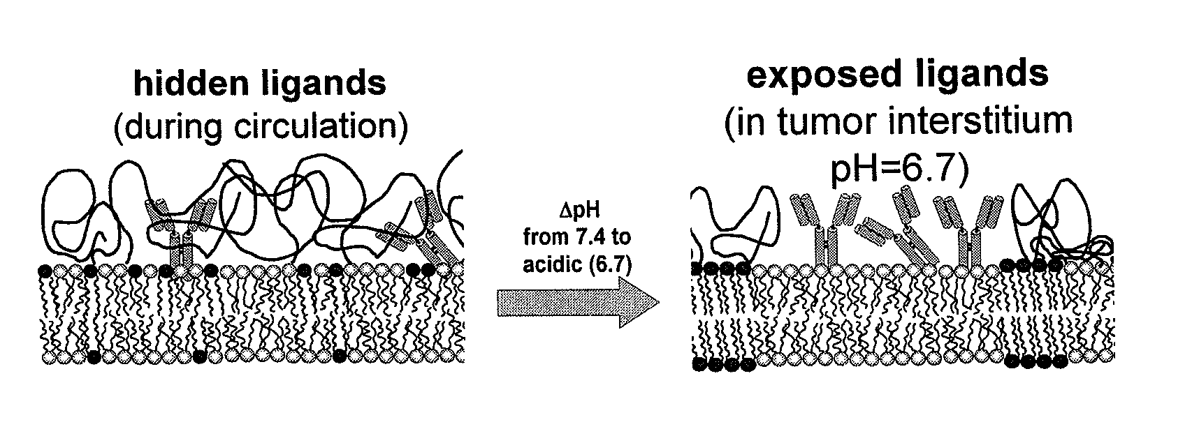
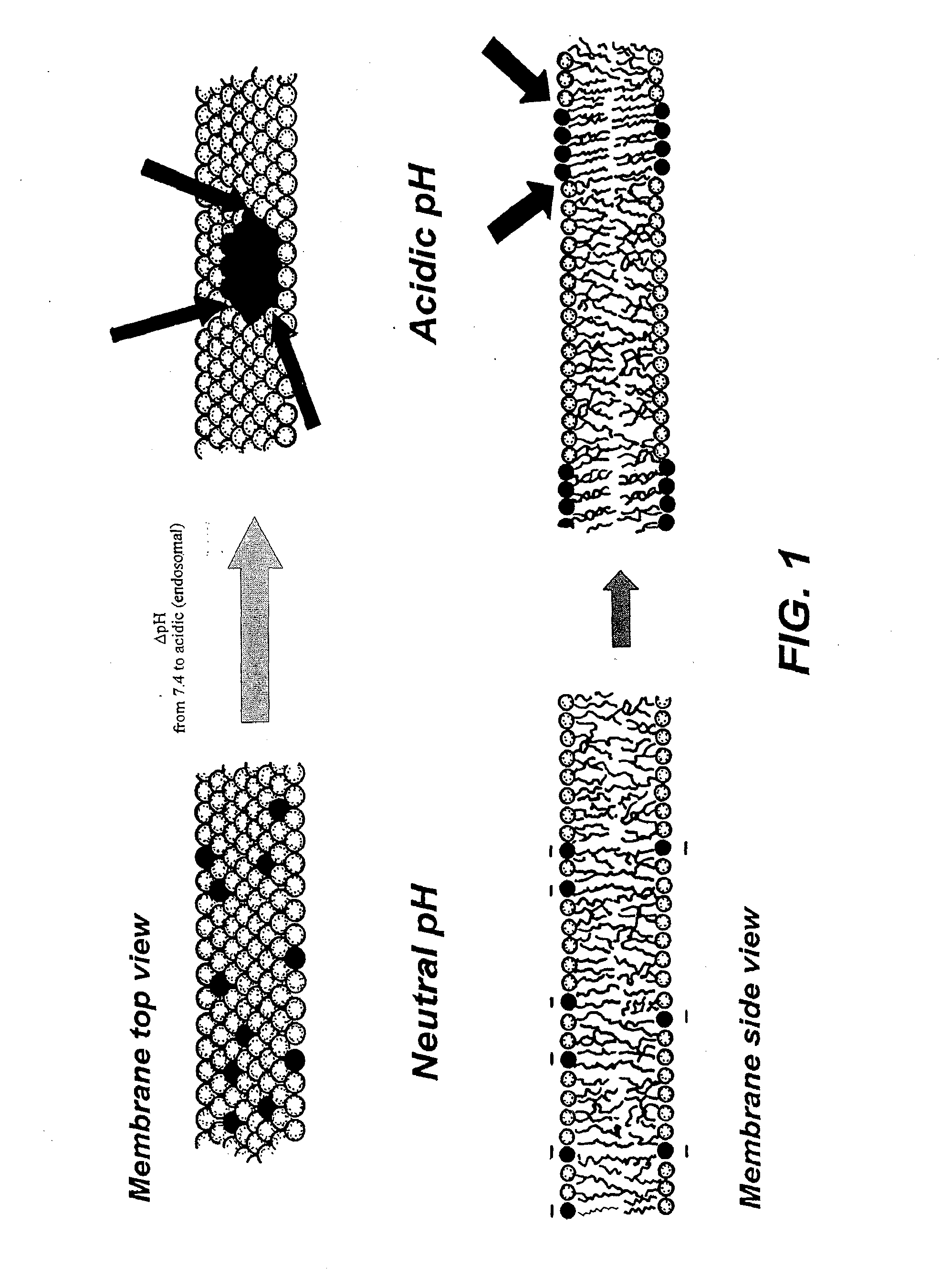
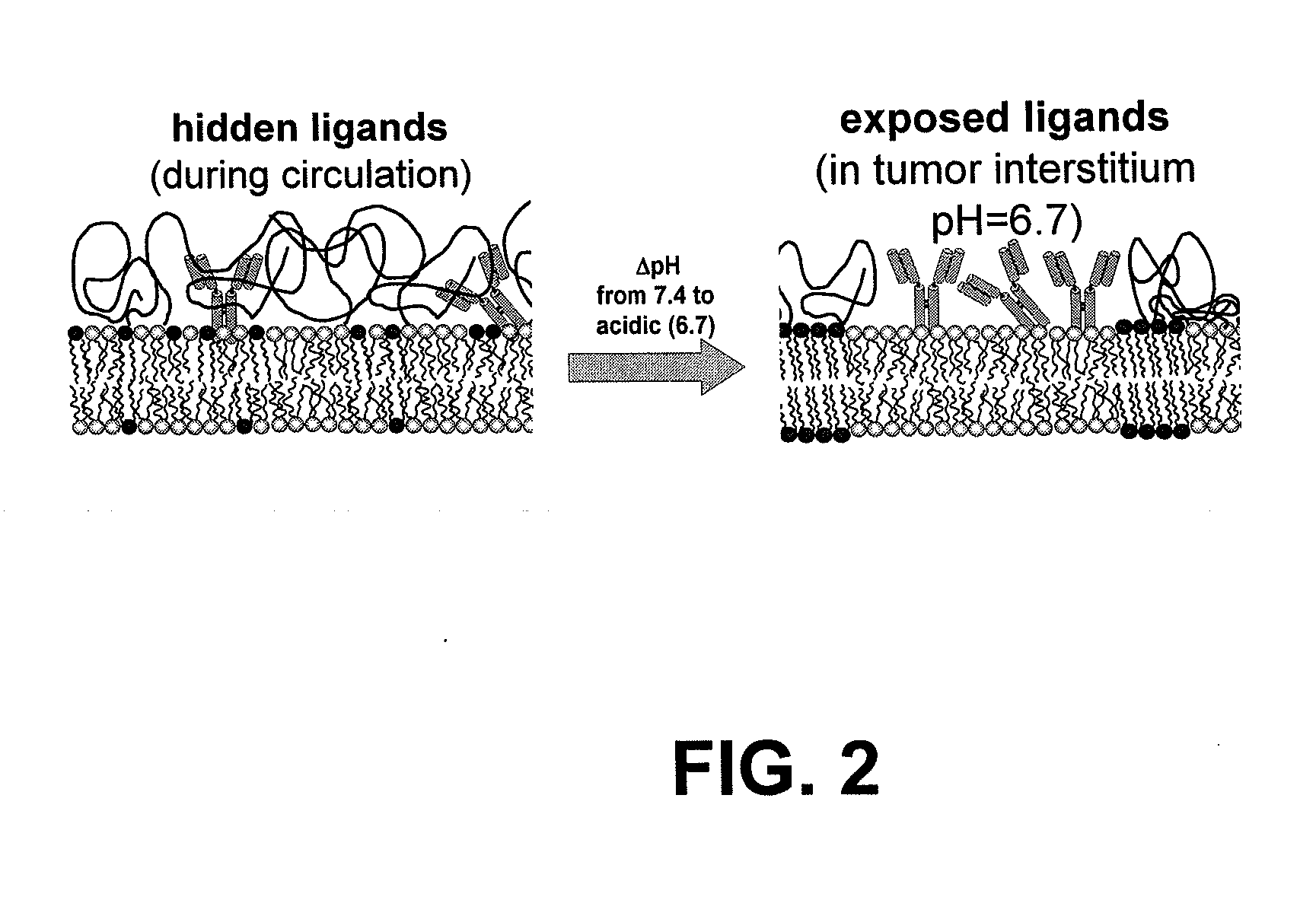
[0082]Biotinylated liposomes (1% mole DPPE-biotin) were developed containing PEGylated lipids that contain domain-forming lipids, which were tuned to become activated at conditions similar to those of tumor interstitial pH. Rigid liposomes consisting of DPPC (16:0), DSPS (18:0) (at 1:1 mole ratios), and 5% cholesterol and 0.1 to 1.5% mole DSPE-PEG (2000 MW) were incubated in PSB at 37° C. at various pH values.

example 2

[0083]Binding of rigid biotinylated liposomes (FIGS. 3, 4, 5, 6, 7, 8, filled symbols) to streptavidin-covered—magnetic microbeads was evaluated at various pH values ranging from pH 7.4, approximating the pH of the blood during circulation of liposomes to pH 6.5 that corresponds to the pH of the tumor interstitium after extravasation of liposomes into the tumor. The extent of liposomes bound was evaluated for different amounts of PEG-linked lipids in the liposome composition ranging from 0.1 to 1.5% mole (of total lipid) and was also compared to identical liposomes without biotin (plain liposomes) indicated by the open symbols in FIGS. 3, 4, 5, 6, 7, and 8. In biotinilated liposomes the amount of biotin-linked lipids was retained constant at 1% mole of total lipid (FIG. 3 shows liposomes containing 0.1% mole PEG-linked lipid, FIG. 4 0.25% mole, FIG. 5 0.5% mole, FIG. 6 0.75% mole, FIG. 7 1.0% mole, and FIG. 8 1.5% mole). The liposomal membrane was labeled with rhodamine, and liposom...

example 3

[0087]Differential Scanning Calorimetry (DSC) was used because it can provide direct evidence of phase separation of lipid membranes. FIG. 11 shows the thermal scans of the same liposome composition (equimolar DPPC and DSPS with 5% mole Cholesterol and 2% mole DSPE-PEG), performed at a rate of 60° C. / h. As the pH was decreased from 7.4 to 4.0, an enhancement was observed on the contributions from thermal transitions at higher temperatures. Higher thermal transitions at lower pH values suggest increasing formation of lipid phases that are rich in clustered (protonated) DSPS lipids (that has higher Tg) and phases poor in DSPS lipids (or richer in DPPC lipids, FIG. 11). These results demonstrate that in membranes containing lipids with different hydrocarbon chain lengths (with one lipid type bearing charged headgroups), lipid mixing or domain formation is controlled by the pH that affects the extent of electrostatic repulsion among the titratable lipids.


PUM

PropertyMeasurementUnit
phase transition temperatureaaaaaaaaaa
pHaaaaaaaaaa
pHaaaaaaaaaa

Abstract

Using liposomes to deliver bioactive agents to cancer or tumor cells and compositions of specific lipids that form liposomes to deliver a biologically active agent.

Description

STATEMENT OF RELATED APPLICATIONS[0001]This application is based on and claims priority on U.S. Provisional Patent Application No. 60 / 828,523 having a filing date of 6 Oct. 2006.BACKGROUND[0002]1. Technical Field[0003]This invention generally relates to the field of therapeutic delivery systems and liposome compositions. Further, this invention is directed to a composition of specific lipids that form liposomes, which can deliver a biologically active agent.[0004]2. Related Art[0005]Lipidic particles can be complexed with virtually any biological material. This capability allows these lipidic particles to be used as delivery systems for bioactive agents. Lipidic complexes have been used for a myriad of drug therapies, and one area in which these delivery systems have shown promising results is in cancer therapies. For a cancer therapy to be successful and efficient, the bioactive agent should be targeted to the tumor or cancer cell.[0006]In some cases, only after the drug carriers a...

Claims


Application Information

Patent Timeline
Patent Type & Authority Applications(United States)
IPC IPC(8): A61K9/127A61K51/12A61K31/7088A61K38/00A61K39/395A61P35/00
CPCA61K9/1271A61P35/00
Inventor STAVROULA, SOFOU
Owner POLYTECHNIC INSTITUTE OF NEW YORK UNIVERSITY
Why Eureka
Try Eureka