Devices and methods for percutaneous access, hemostasis, and closure

a technology of percutaneous access and hemostasis, applied in the field of devices and methods for percutaneous access, hemostasis, and closure, can solve the problems of minimally invasive approaches, introduce new complexities of surgery, and minimally invasive approaches, and achieve the effect of high-profile configuration

Inactive Publication Date: 2011-02-17
NORTHWESTERN UNIV
View PDF16 Cites 118 Cited by
  • Summary
  • Abstract
  • Description
  • Claims
  • Application Information

AI Technical Summary

Problems solved by technology

The use of minimally invasive approaches, however, introduces new complexities to surgery.
An inherent difficulty in the minimally invasive percutaneous approach is the limited space that is available within the vasculature.
Consequently, the introduction of tools and prosthetic devices becomes a great deal more complicated as compared to open-chest surgeries.
One route of access, through the femoral arteries, is limited because of vascular disease involving these vessels and the size of the devices to be placed is largely relative to the small diameter vessels.
Transapical placement, through the apex of the left ventricle, has been the preferred approach in many patients because there are less vascular complications, but there are problems such as bleeding from the left ventricular apex requiring difficult suture closure, and a thoracotomy incision that can be painful and lead to difficult recovery and prolonged hospital stay for the patient.

Method used


Image

  • Devices and methods for percutaneous access, hemostasis, and closure
  • Devices and methods for percutaneous access, hemostasis, and closure
  • Devices and methods for percutaneous access, hemostasis, and closure

Examples


example 1

Deployment of A Medical Closure Device of the Present Invention

[0046]The following example provides an exemplary deployment procedure for an exemplary medical closure device. This example should not be viewed as limiting, as deployment procedures, delivery components, and medical devices may differ from the example presented herein.

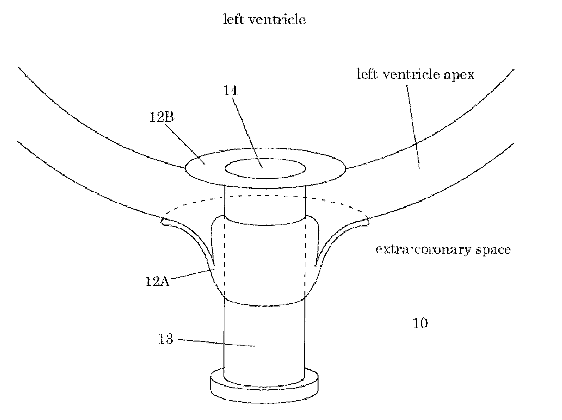
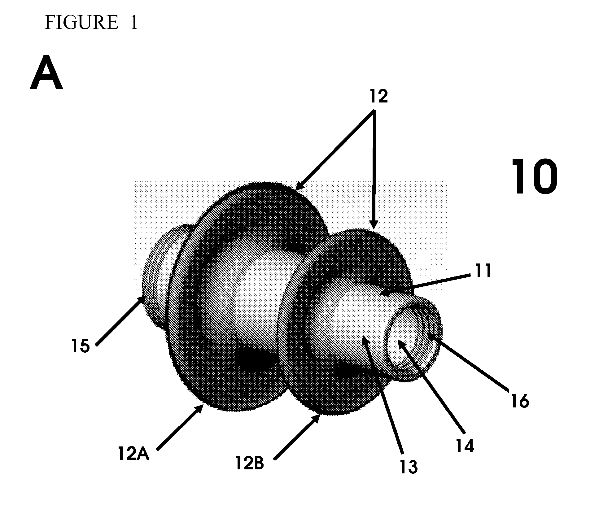
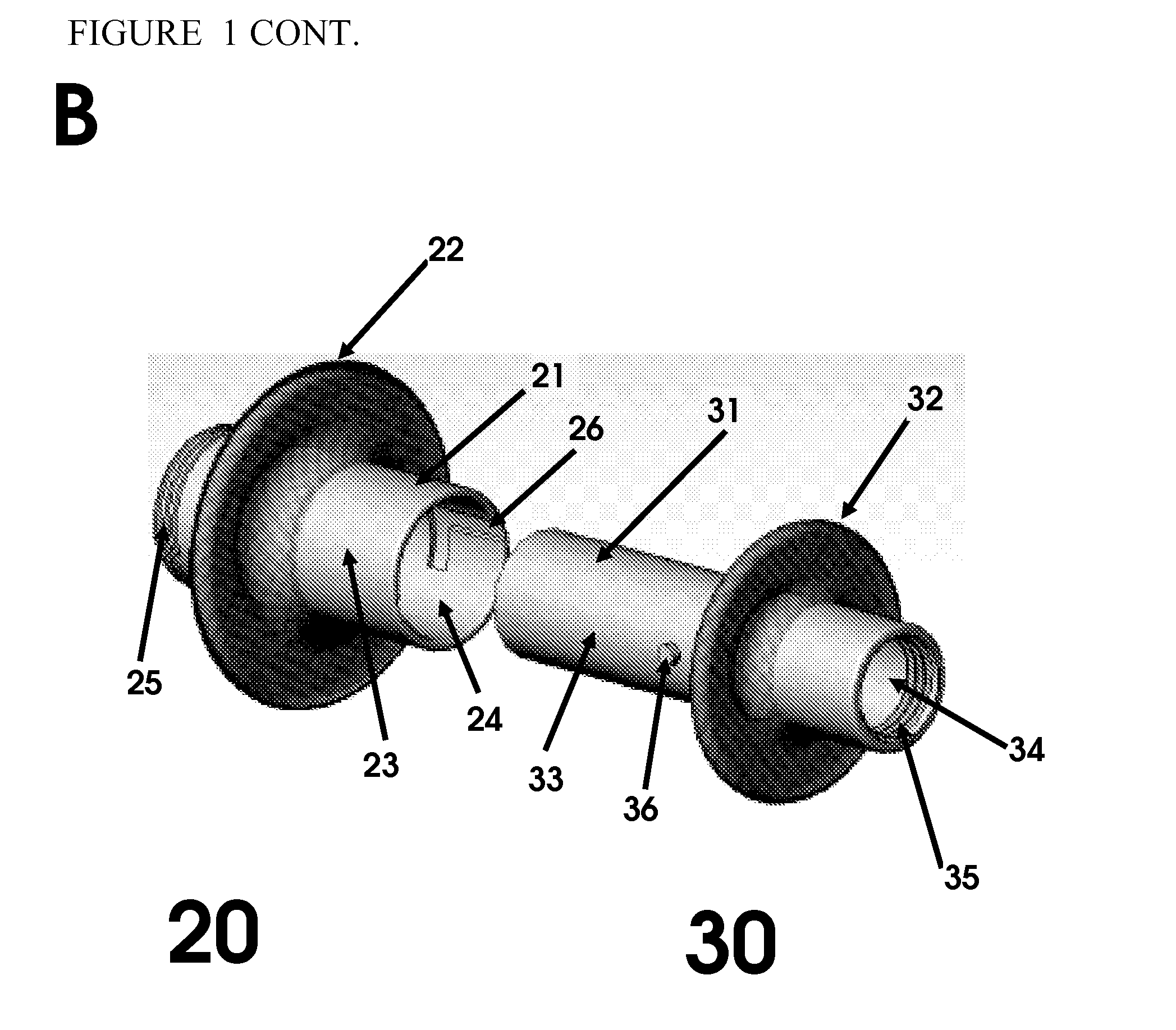
[0047]A medical closure device is placed into the lumen of a delivery catheter and positioned at the distal end of the catheter. The distal end of the medical closure device is positioned facing outward from the distal end of the delivery catheter. The confined space of the lumen of the delivery catheter compresses the distal and proximal expanded elements causing the expanded elements to adopt a reduced cross-sectional profile configuration.

[0048]The delivery catheter is deployed via a minimally invasive pathway through a rib space directed toward the ventricular apex of the heart of a human patient (SEE FIG. 4A). The delivery catheter is positioned upon...

example 2

Access, Hemostasis, And Closure

[0050]The following example provides exemplary methods of use for an exemplary medical closure device. This example should not be viewed as limiting, as medical closure devices of the present invention find utility in a range of procedures and surgeries not detailed in the following example.

[0051]A medical closure device is deployed during a medical procedure to provide access to the procedure site. The device is deployed, as described in Example 1 for example, to the left ventricular apex of the heart of a patient. Upon insertion into the apical tissue, the device provides an access port for performing medical and surgical procedures within the left ventricular space. A one-way valve within the inner lumen of the medical closure device prevents the outflow of blood and fluid from the left ventricle or other heart chambers into the extra-cardiac space. The valve further provides access to the chambers of the heart for surgical and medical instruments. ...


PUM


Abstract

The present invention provides methods and devices for obtaining percutaneous access, maintaining hemostasis, and providing closure of openings in tissues of the body. In particular, the present invention provides methods and devices for maintaining hemostasis and providing closure surgical incisions of the left ventricular apex of the heart.

Description

[0001]The present application claims priority to U.S. Provisional Patent Application Ser. No. 61 / 037,255 filed Mar. 17, 2008, the entire disclosure of which are herein incorporated by reference in its entirety.FIELD OF THE INVENTION[0002]The present invention provides methods and devices for obtaining percutaneous access, maintaining hemostasis, and providing closure of openings in tissues of the body. In particular, the present invention provides methods and devices for maintaining hemostasis and providing closure surgical incisions of the left ventricular apex of the heart.BACKGROUND OF THE INVENTION[0003]Historically, human heart valves have been replaced using open heart surgery involving the heart / lung machine and excision of the native aortic valve and suture placement of the new valve prosthesis. New technology has been developed to enable valve replacement “percutaneously,” or through a “transapical” procedure, using a device with a valve inside a stent placed within the pat...

Claims


Application Information

Patent Timeline
Patent Type & Authority Applications(United States)
IPC IPC(8): A61B17/12
CPCA61B17/3421A61B17/3423A61B2017/00243A61B2017/00252A61B2017/00575A61B2017/3425A61B2017/0237A61B2017/3488A61B17/0057A61B17/0218A61B2017/00606A61B2017/00867A61B2017/3484
Inventor MCCARTHY, PATRICK M.GNANASHANMUGAM, SWAMINADHANPERIYANAYAGAM, USHALIN, INGRIDRALPH, LEAHLUBECK, CHRISTOPHER
Owner NORTHWESTERN UNIV
Why Eureka
Try Eureka