
Magnetic resonance imaging apparatus and magnetic resonance imaging method

Active Publication Date: 2011-03-31
TOSHIBA MEDICAL SYST CORP
View PDF5 Cites 20 Cited by
  • Summary
  • Abstract
  • Description
  • Claims
  • Application Information

AI Technical Summary

Benefits of technology

[0016]The present invention aims to provide a technology capable of imaging an image allowing a dynamic behavior of CSF to be better understood without using a contrast medium in magnetic resonance imaging.
[0023]According to the aforementioned magnetic resonance imaging of the present invention, it is possible to acquire a CSF image allowing a dynamic behavior of CSF to be better understood without using a contrast medium.

Problems solved by technology

In light of this, from images acquired by the t-SLIP method, it is possible to understand a dynamic behavior of a periodic fluid, but it is difficult to understand a dynamic behavior of a non-periodic fluid accurately.
For this reason, the method of applying a plurality of labeling pulses cannot generate images representing a dynamic fluid behavior of synchronous time.
In light of this, the t-SLIP method of performing data acquisition a plurality of times by changing the BBTI has difficulty in following and imaging the CSF whose flow is changed.
For this reason, the t-SLIP method cannot allow a wide range of CSF flow of synchronous time to be understood.
For this reason, in the t-SLIP method, it is very difficult to visually understand a detailed dynamic CSF behavior from monochrome image in grayscale.

Method used


Image

  • Magnetic resonance imaging apparatus and magnetic resonance imaging method
  • Magnetic resonance imaging apparatus and magnetic resonance imaging method
  • Magnetic resonance imaging apparatus and magnetic resonance imaging method

Examples


Embodiment Construction

[0040]A magnetic resonance imaging apparatus and a magnetic resonance imaging method according to embodiments of the present invention will be described with reference to the accompanying drawings.

(Configuration and Function)

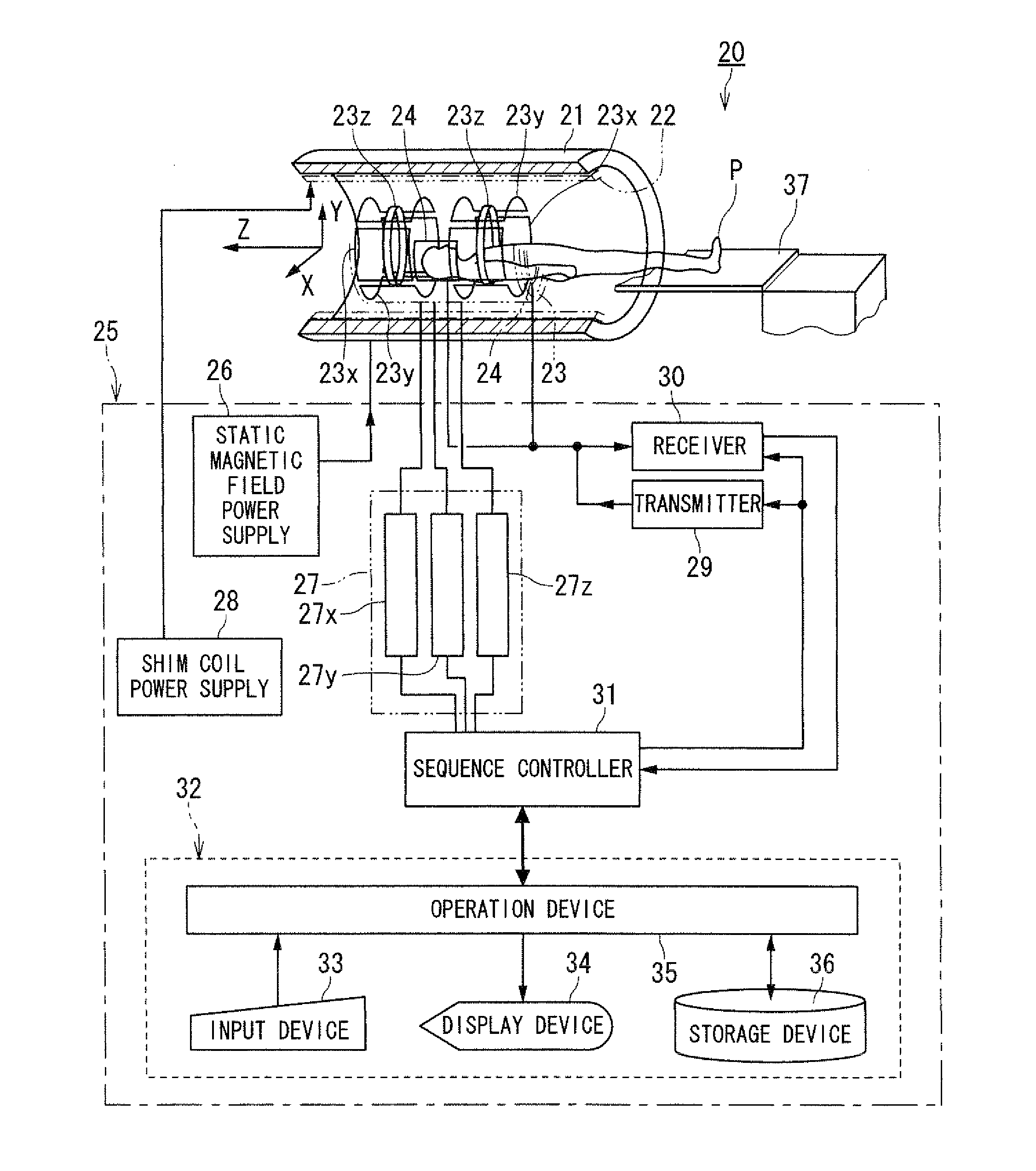
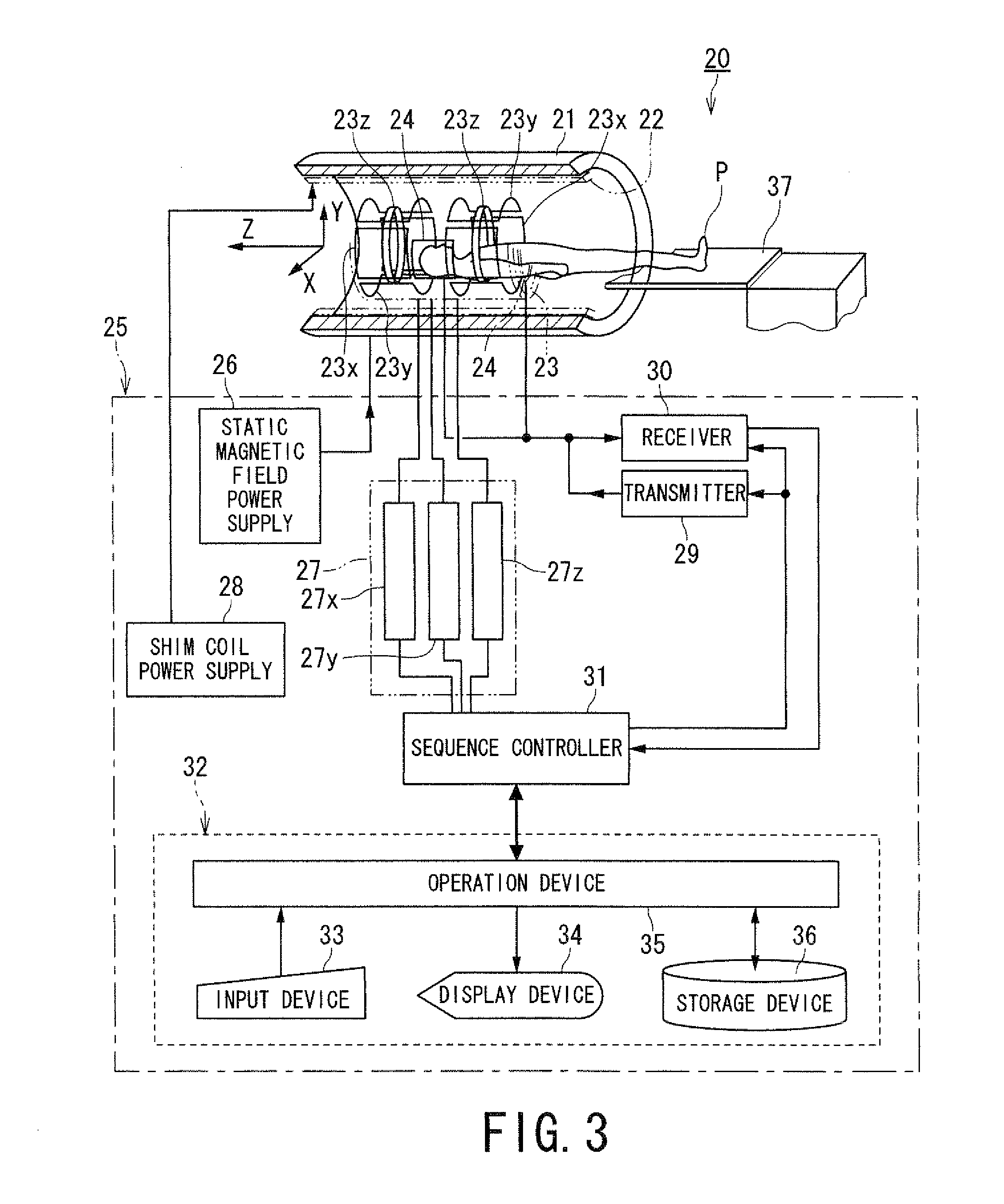
[0041]FIG. 3 is a block diagram showing a magnetic resonance imaging apparatus according to an embodiment of the present invention.

[0042]A magnetic resonance imaging apparatus 20 includes a cylinder-shaped static field magnet 21 for generating a static magnetic field, a cylinder-shaped shim coil 22 arranged inside the static field magnet 21, a gradient coil 23 and RF coils 24.

[0043]The magnetic resonance imaging apparatus 20 also includes a control system 25. The control system 25 includes a static magnetic field power supply 26, a gradient magnetic field power supply 27, a shim coil power supply 28, a transmitter 29, a receiver 30, a sequence controller 31 and a computer 32. The gradient magnetic field power supply 27 of the control system 25 includes an X-axis...


PUM


Abstract

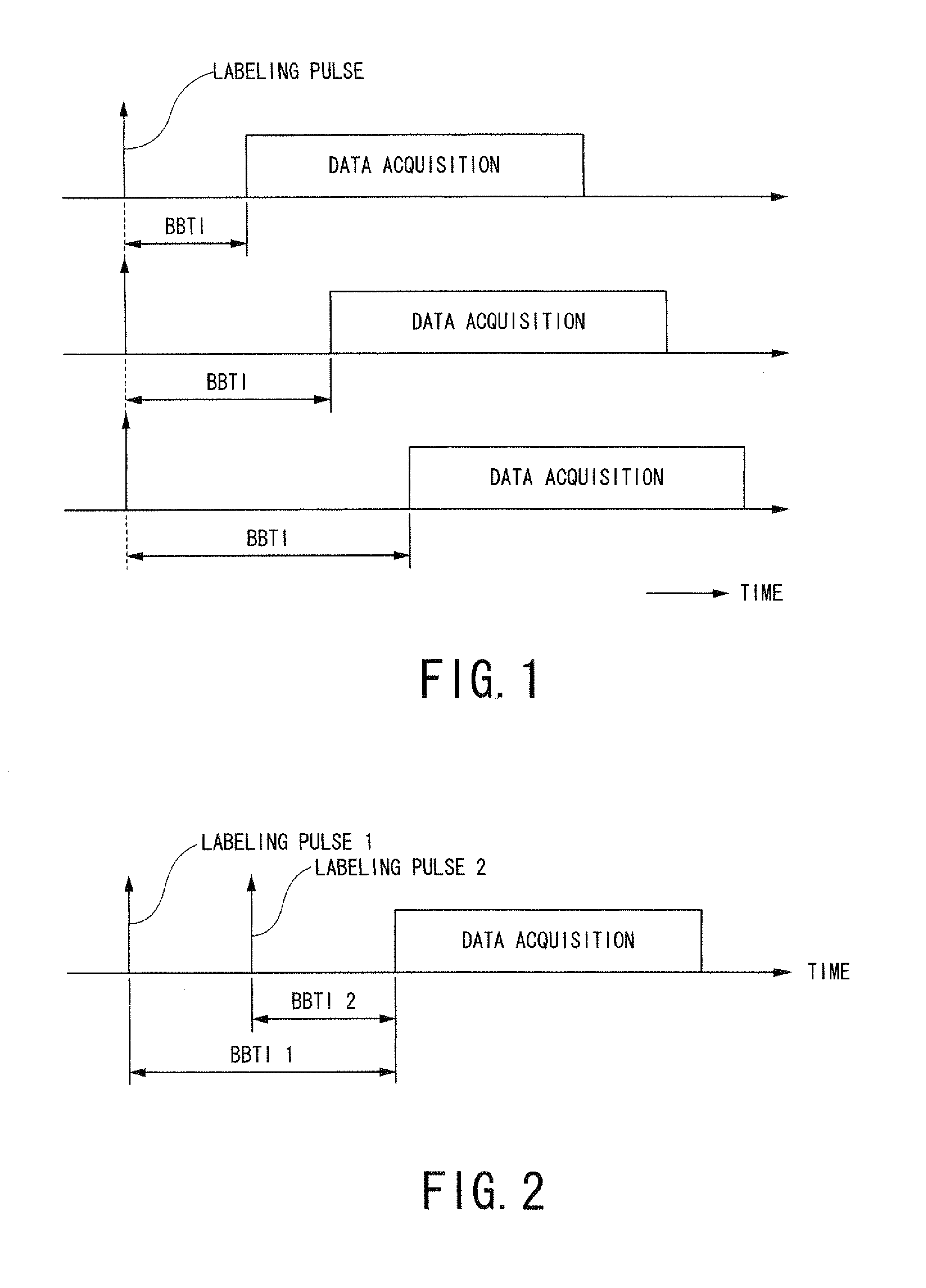
A magnetic resonance imaging apparatus includes a magnetic resonance data acquisition unit and a cerebrospinal fluid image data generation unit.The magnetic resonance data acquisition unit consecutively acquires a plurality of magnetic resonance data for generating a plurality of cerebrospinal fluid image data, each corresponding to a different data acquisition time, after a labeling pulse is applied.The cerebrospinal fluid image data generation unit generates the plurality of cerebrospinal fluid image data based on the plurality of magnetic resonance data.

Description

BACKGROUND OF THE INVENTION[0001]1. Field of the Invention[0002]The present invention relates to MRI (magnetic resonance imaging) which excites nuclear spin of an object magnetically with an RF (radio frequency) signal having the Larmor frequency and reconstruct an image based on NMR (nuclear magnetic resonance) signals generated due to the excitation. More particularly, the present invention relates to a magnetic resonance imaging apparatus and a magnetic resonance imaging method which can image CSF (cerebrospinal fluid).[0003]2. Description of the Related Art[0004]Magnetic Resonance Imaging is an imaging method which excites nuclear spin of an object set in a static magnetic field with an RF signal having the Larmor frequency magnetically and reconstruct an image based on NMR signals generated due to the excitation.[0005]In the field of the magnetic resonance imaging, MRA is known as a method of obtaining an image of a blood flow. An MRI without using a contrast medium is referred...

Claims


Application Information

Patent Timeline
IPC IPC(8): G01R33/48
CPCA61B5/055A61B5/7285G01R33/5602A61B5/0042G01R33/54G01R33/28G06T7/0012G01R33/56333
Inventor YAMASHITA, YUICHIICHINOSE, NOBUYASUKITANE, SHINICHI
Owner TOSHIBA MEDICAL SYST CORP
Learn More