Magnetic medical apparatus, kits, and methods

a magnetic medical device and kit technology, applied in the field of medical devices, can solve the problems of limiting the inner diameter of conventional vascular grafts to 6 millimeters, and achieve the effects of convenient coating of medical devices, easy magnetization, and rapid attraction and attachmen

Active Publication Date: 2011-05-19
MAYO FOUND FOR MEDICAL EDUCATION & RES
View PDF61 Cites 7 Cited by
  • Summary
  • Abstract
  • Description
  • Claims
  • Application Information

AI Technical Summary

Benefits of technology

[0019]One potential advantage of the present invention is the ability to rapidly attract and attach large numbers of magnetic cells and other magnetic materials to the magnetic contact surfaces of a medical device. This is possible because the device can be located in a carrier liquid containing high concentrations of magnetic cells / material before or after implantation. The carrier liquid with magnetic cells / material may be in contact with the medical device in vitro or in vivo. In either case, it may be preferred that the carrier liquid have a concentration of magnetic cells / material that is high enough to facilitate coating of the medical device within an acceptable period of time, e.g., several hours or less.
[0020]Although described herein in the context of attracting magnetic cells, it should be understood that the medical devices, methods, and kits of the present invention may be used to attract magnetic materials other than cells, e.g., drugs, therapeutic substances, sub-cellular particles, etc.
[0021]If the magnetic coating is performed in vivo, it may be preferred that the carrier liquid with magnetic cells be delivered to a defined volume in which the medical device is located. If the medical device is implanted within, e.g., a blood vessel, the defined volume may be created by using a catheter to seal the vessel on one or both sides of the implanted medical device, followed by delivery of the carrier liquid to the location of the implanted medical device.
[0022]Another potential advantage of the present invention is that the medical devices may be located in any suitable location within the body and coated with magnetic cells after implantation, i.e., the medical devices and methods may be used in blood vessels, but the potential locations are not limited to blood vessels. For example, the medical devices may be located and magnetically coated within the gastrointestinal tract, salivary glands, bile ducts, pancreatic ducts, renal system (kidney, ureters, bladder, urethra, etc.), airways in lungs, conduits in the brain and spinal cord, surgically created conduits or lumens in an organ, congenital abnormalities in any organ, etc.
[0023]Another potential advantage of the present invention is that medical devices constructed of metals and / or metal alloys may be easily and quickly magnetized by exposure to a magnetic field (if the metals or metal alloys do not inherently exhibit magnetic fields). In some embodiments, the apparatus and methods of the present invention provide the ability to magnetize a medical device as part of the implantation process or to magnetize the medical device in vivo. In some instances, it may be possible to use known medical devices such as, e.g., stents, heart valves, etc. in connection with the present invention.
[0024]Yet another potential advantage of the present invention is the ability to demagnetize an implanted medical device after magnetically coating the medical device by, e.g., locating a degaussing element proximate the medical device in vivo after a selected period of time, e.g., 24 hours to 48 hours. By magnetically coating the medical device, the advantages associated with magnetic coating as discussed herein may be obtained and any potential disadvantages associated with leaving a magnetized medical device may be alleviated by demagnetizing the medical device after the magnetically attracted biologic cells have formed alternative attachments to the medical device.

Problems solved by technology

Conventional vascular grafts are typically limited to inner diameters of 6 millimeters (mm) or more because of occlusion problems.

Method used


Image

  • Magnetic medical apparatus, kits, and methods
  • Magnetic medical apparatus, kits, and methods
  • Magnetic medical apparatus, kits, and methods

Examples


Embodiment Construction

[0073]In the following detailed description of illustrative embodiments, reference is made to the accompanying drawings that form a part hereof, and in which are shown, by way of illustration, specific embodiments in which the invention may be practiced. It is to be understood that other embodiments may be utilized and structural changes may be made without departing from the scope of the present invention. Furthermore, like reference numbers denote like features in the different figures.

[0074]It should be noted that as used herein and in the appended claims, the singular forms “a”, “and”, and “the” include plural referents unless the context clearly dictates otherwise. Thus, for example, reference to “a magnetic cell” includes a plurality of such cells and reference to “the magnetic contact surface” includes reference to one or more magnetic contact surfaces and equivalents thereof known to those skilled in the art.

Medical Devices with Magnetic Contact Surfaces

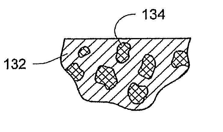
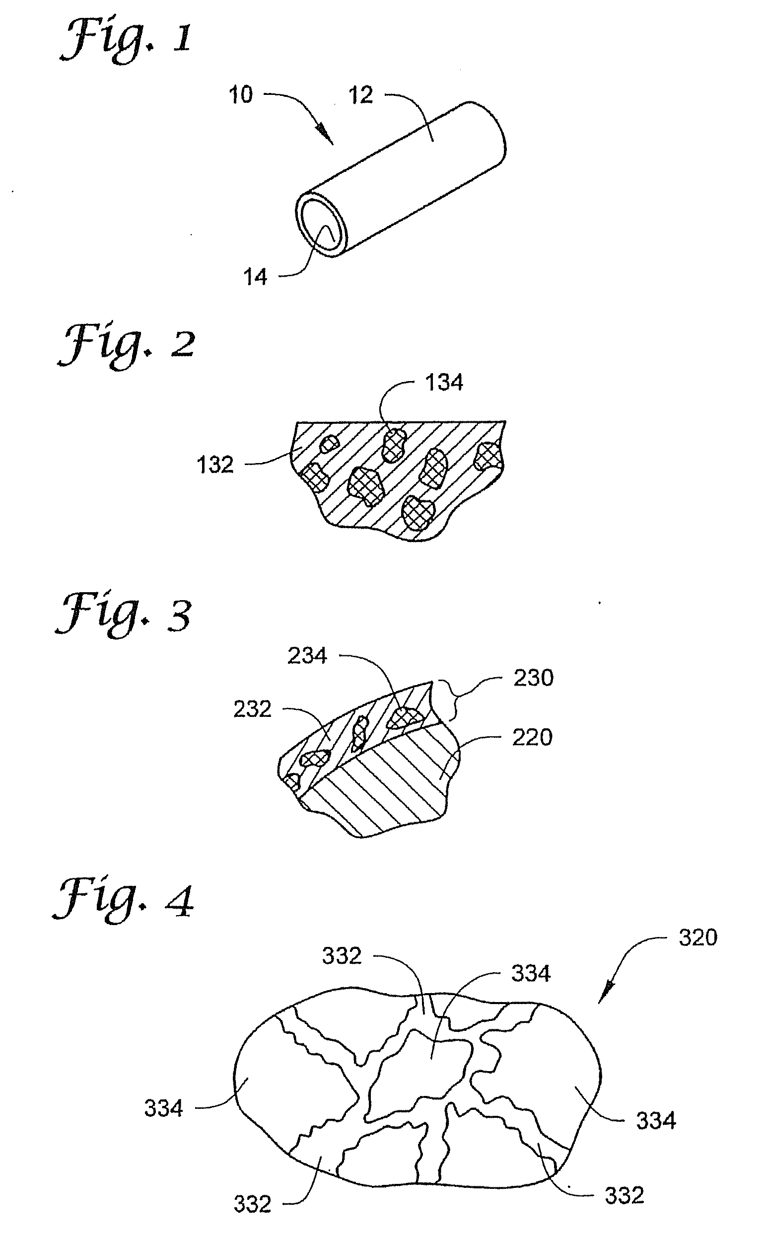
[0075]FIG. 1 depicts ...


PUM


Abstract

Medical devices with surfaces on which viable biologic cells are magnetically attracted and retained are disclosed along with methods of magnetic coating. The medical devices can be located in a carrier liquid containing high concentrations of magnetic cells before or after implantation. The carrier liquid with magnetic cells may be contact with the medical device in vitro or in vivo. In either case, the carrier liquid may have a concentration of magnetic cells that is high enough to facilitate coating of the medical device within an acceptable period of time, e.g., several hours or less. Magnetization of medical devices before, during, and / or after implantation and apparatus for performing the same are disclosed. Degaussing of magnetic medical devices is also disclosed.

Description

RELATED APPLICATIONS[0001]This application claims the benefit of U.S. Provisional Application No. 60 / 691,090, filed on 16 Jun. 2005, titled MEDICAL DEVICES WITH DISTRIBUTED MAGNETIC FIELDS AND METHODS OF MANUFACTURING THE SAME, which is hereby incorporated by reference in its entirety.[0002]This application is also a continuation-in-part of U.S. patent application Ser. No. 11 / 446,861, filed on 5 Jun. 2006, titled KITS, APPARATUS AND METHODS FOR MAGNETICALLY COATING MEDICAL DEVICES WITH LIVING CELLS, which claims the benefit of U.S. Provisional Application No. 60 / 691,090, filed on 16 Jun. 2005, titled MEDICAL DEVICES WITH DISTRIBUTED MAGNETIC FIELDS AND METHODS OF MANUFACTURING THE SAME and which is a continuation-in-part of PCT / US2004 / 040242, filed on 2 Dec. 2004, titled KITS, APPARATUS AND METHODS FOR MAGNETICALLY COATING MEDICAL DEVICES WITH LIVING CELLS, which claims the benefit of U.S. Provisional Application No. 60 / 526,522, filed on 3 Dec. 2003, titled COATING MAGNETIC MEDICAL ...

Claims


Application Information

Patent Timeline
Patent Type & Authority Applications(United States)
IPC IPC(8): A61F2/82A61F2/04A61F2/06
CPCA61F2/04A61F2/06A61N2001/0585A61F2/82A61F2210/009A61F2/24A61F2/07A61F2/30A61F2002/077A61F2002/30079
Inventor SANDHU, GURPREET S.SIMARI, ROBERT D.SANDHU, NICOLE P.GULATI, RAJIV
Owner MAYO FOUND FOR MEDICAL EDUCATION & RES
Why Eureka
Try Eureka