Ultrasonic diagnostic apparatus and method for calculating coordinates of scanned surface thereof

a diagnostic apparatus and ultrasonic technology, applied in diagnostics, medical science, applications, etc., can solve the problems of difficult time-consuming, difficult to set the same conditions for the positional relationship of objects, and difficult to compare and display the same cross-section in tomographic images of objects in pre-treatment and post-treatment, so as to facilitate match the display cross-section

Inactive Publication Date: 2011-06-16
HITACHI MEDICAL CORP
View PDF5 Cites 17 Cited by
  • Summary
  • Abstract
  • Description
  • Claims
  • Application Information

AI Technical Summary

Benefits of technology

[0023]In this manner, since coordinates of the scanned plane of the real-time ultrasonic tomographic image can be calculated in association with the object coordinate system at the time of storing the data even when the object coordinate system at the time of storing the data and the object coordinate system at the time of comparing the images are different, the display cross-section of the earlier-scanned tomographic image can be easily matched with the display cross-section of the later-scanned tomographic image.
[0024]Thus, in accordance with the present invention, it is possible to easily match the display cross-sections of the earlier-scanned tomographic image and the later-scanned tomographic image.

Problems solved by technology

However, it generally is difficult to set the same conditions for the positional relationship of the object, etc. each time pre-treatment images and post-treatment images are scanned.
Given this factor, it is necessary to estimate the imaging position of the pre-treatment image referring to the stored ultrasonic tomographic images, but comparing and displaying the same cross-sections in the tomographic images of an object in pre-treatment and post-treatment is difficult and time consuming.

Method used


Image

  • Ultrasonic diagnostic apparatus and method for calculating coordinates of scanned surface thereof
  • Ultrasonic diagnostic apparatus and method for calculating coordinates of scanned surface thereof
  • Ultrasonic diagnostic apparatus and method for calculating coordinates of scanned surface thereof

Examples


embodiment 1

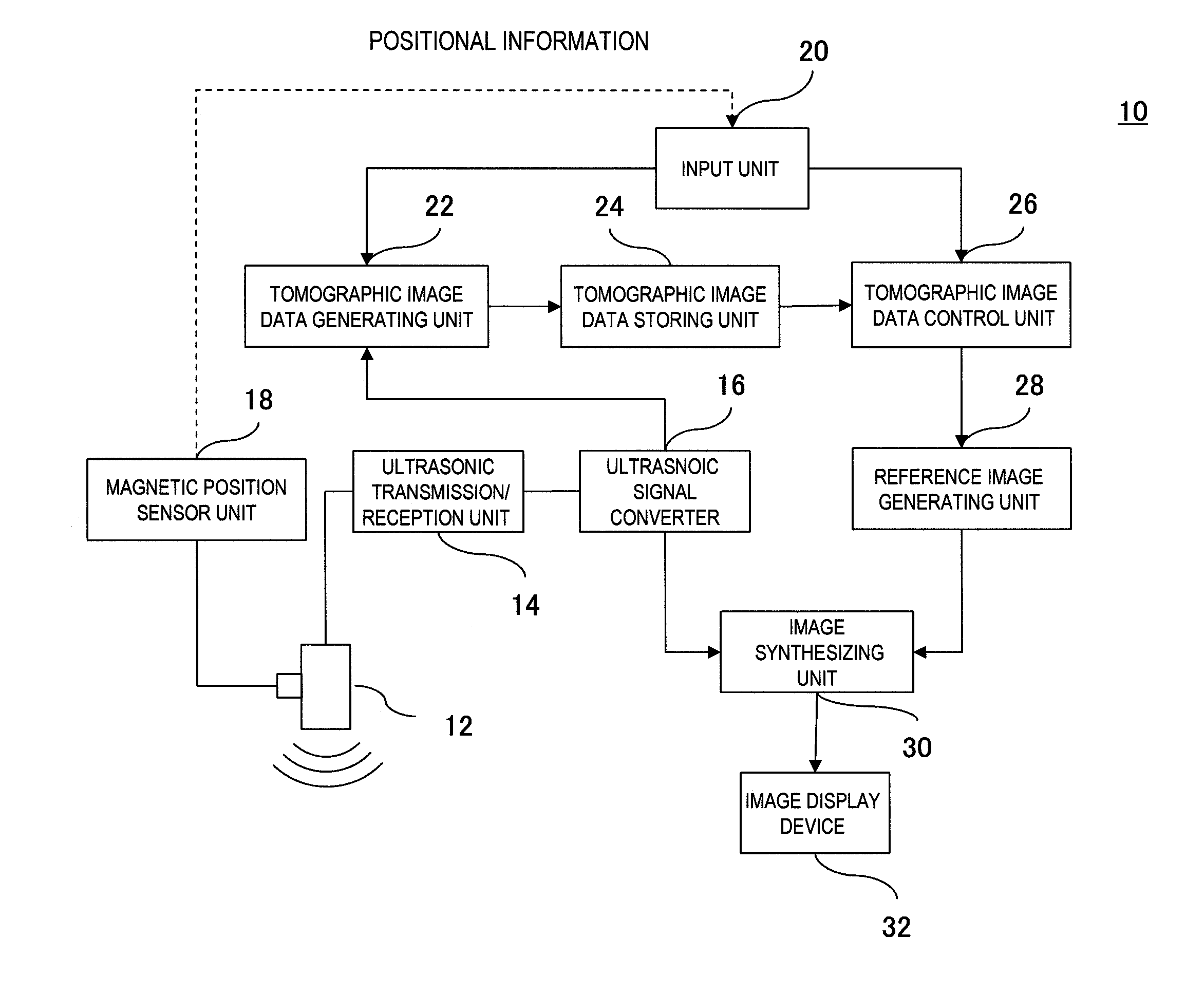
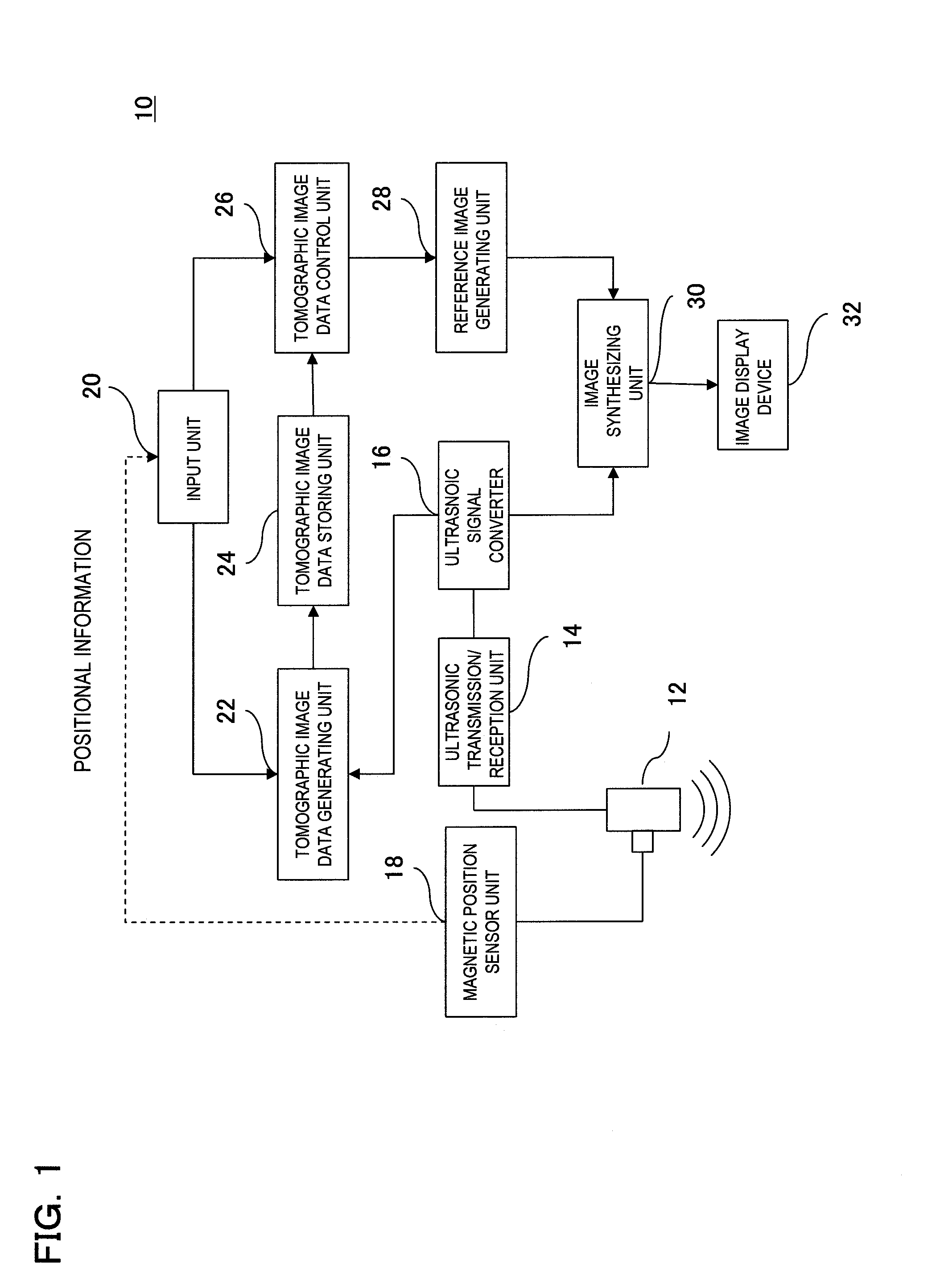
[0053]The present embodiment stores the 3-dimensional volume data formed by plural sets of ultrasonic tomographic image data in a storage means, extracts the ultrasonic tomographic image of the same cross-section as the present ultrasonic scanned plane from the stored 3-dimensional volume data, and displays the extracted image along with the real-time ultrasonic tomographic image.

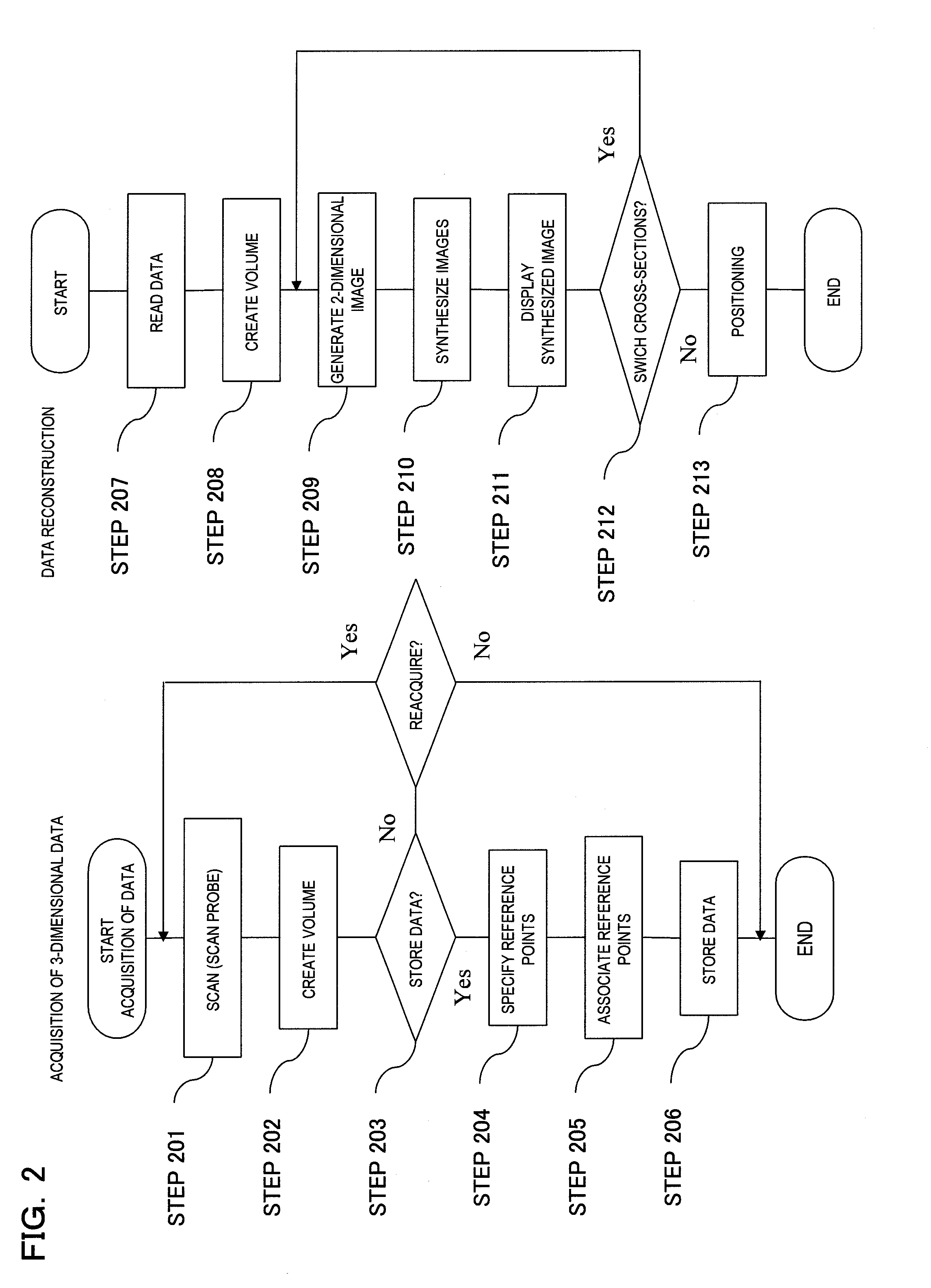
[0054]FIG. 2 is a flowchart showing acquisition, storage and reconstruction process of the 3-dimensional volume data in the present embodiment. FIG. 3 shows a schematic diagram and display example, etc. in each process of the present embodiment.

[0055]As shown in FIG. 3A, the body surface of object 40 is scanned by ultrasonic probe 12 to which a magnetic position sensor is attached, consecutive 2-dimensional ultrasonic tomographic images are obtained (step 201), and the 3-dimensional volume data is generated from a plurality of 2-dimensional tomographic images (step 202). In the case of storing the 3-dimensi...

embodiment 2

[0073]In the present embodiment, not 3-dimensional volume data formed by plural sets of ultrasonic tomographic data but one set or plural sets of ultrasonic tomographic data which do not construct 3-dimensional volume data are stored in storage means, and position of an ultrasonic probe is guided for displaying the real-time ultrasonic tomographic image of the same cross-section as the stored ultrasonic tomographic image data. The difference from the first embodiment will be mainly described below and the duplicative description thereof will be appropriately omitted.

[0074]FIG. 5 is a flowchart of acquisition, storage and reconstruction process of 2-dimensional ultrasonic tomographic image data in the present embodiment. FIG. 6 shows schematic diagrams, display example, etc. in each process of the present embodiment.

[0075]First, discriminative three points are specified on body contour model 42 by the same operation in the first embodiment (step 501), and the body contour model and t...


PUM


Abstract

An ultrasonic diagnostic apparatus includes a storage unit configured to store information about the contour of an object to be examined and information about the position of a specified region of the ultrasonic image of the object both corresponded to a body contour model coordinate system and a coordinate calculating unit configured to correspond the object coordinate system to the body contour model coordinate system according to the information about the position of the specified region of the object, associating with each other the object coordinate system and the body contour model coordinate system used when comparing an ultrasonic tomographic image based on ultrasonic tomographic data stored in an image storage unit with a real-time ultrasonic tomographic image scanned by an ultrasonic probe, and thereby calculating the coordinates of the scanned plane of the real-time ultrasonic tomographic image in association with the object coordinate system.

Description

FIELD OF THE INVENTION[0001]The present invention relates to an ultrasonic diagnostic apparatus and method of calculating coordinates of the scanned surface thereof, in particular to a technique for making diagnosis by comparing two of the same cross-sectional images of an object that are imaged at different times.DESCRIPTION OF RELATED ART[0002]An ultrasonic diagnostic apparatus is for making diagnosis of an imaged region noninvasively and in real time by transmitting / receiving ultrasonic waves to / from an object to be examined via a probe, reconstructing and displaying a tomographic image of the imaged region based on the reflected echo signals outputted from the probe.[0003]Such ultrasonic diagnostic apparatus makes diagnosis by displaying and comparing, for example a tomographic image of a diseased area imaged before treatment (hereinafter referred to as a pre-treatment image) and a tomographic image of the diseased area imaged during or after the treatment (hereinafter referred ...

Claims


Application Information

Patent Timeline
Patent Type & Authority Applications(United States)
IPC IPC(8): A61B8/14
CPCA61B8/4254A61B8/14
Inventor NIHEI, KOICHISHINOHARA, DAI
Owner HITACHI MEDICAL CORP
Why Eureka
Try Eureka