Handheld force-controlled ultrasound probe

a force-controlled, ultrasound probe technology, applied in tomography, instruments, applications, etc., can solve the problems of deformation of the internal structure of the patient's tissue, difficulty in medical diagnosis, and difficulty in determining whether the tumor has grown, so as to reduce the force differential

Inactive Publication Date: 2011-07-14
MASSACHUSETTS INST OF TECH
View PDF11 Cites 11 Cited by
  • Summary
  • Abstract
  • Description
  • Claims
  • Application Information

AI Technical Summary

Benefits of technology

[0009]A method disclosed herein includes identifying a predetermined, desired contact force between the handheld ultrasound probe and a target; measuring a current contact force between the handheld ultrasound probe and the target; compar

Problems solved by technology

However, for soft areas of the body, varying the contact force of the ultrasound probe also tends to deform the internal structure of the patient's tissue.
This may lead to difficulties in medical diagnosis, such as where a tumor in one ultrasound image appears to be larger than the same tumor shown in an earlier-acquired image.
It may be difficult or impossible to determine whether the tumor

Method used


Image

  • Handheld force-controlled ultrasound probe
  • Handheld force-controlled ultrasound probe
  • Handheld force-controlled ultrasound probe

Examples


Embodiment Construction

lude two plates positioned to surround and securely affix the handheld ultrasound probe to the substantially rigid frame.

BRIEF DESCRIPTION OF THE FIGURES

[0016]The invention and the following detailed description of certain embodiments thereof may be understood by reference to the following figures:

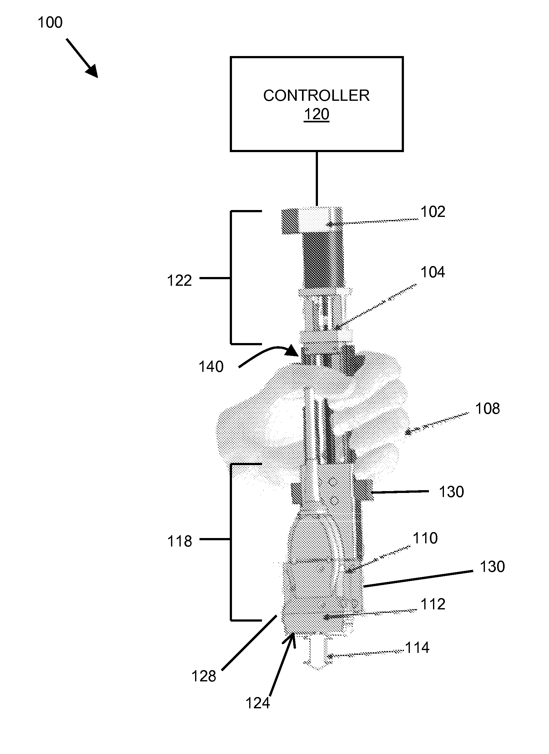
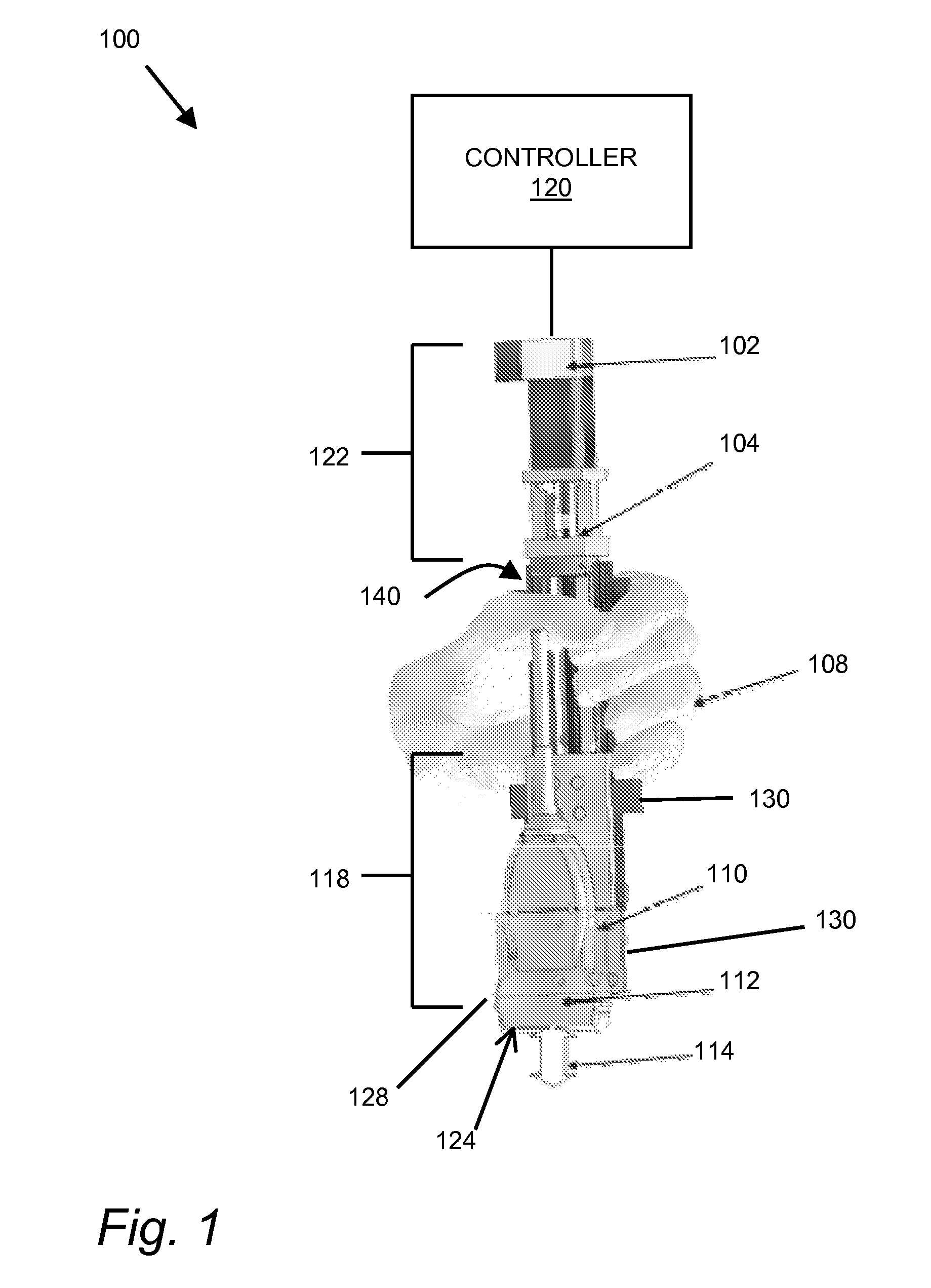
[0017]FIG. 1 is a perspective view of a handheld ultrasound probe control device.

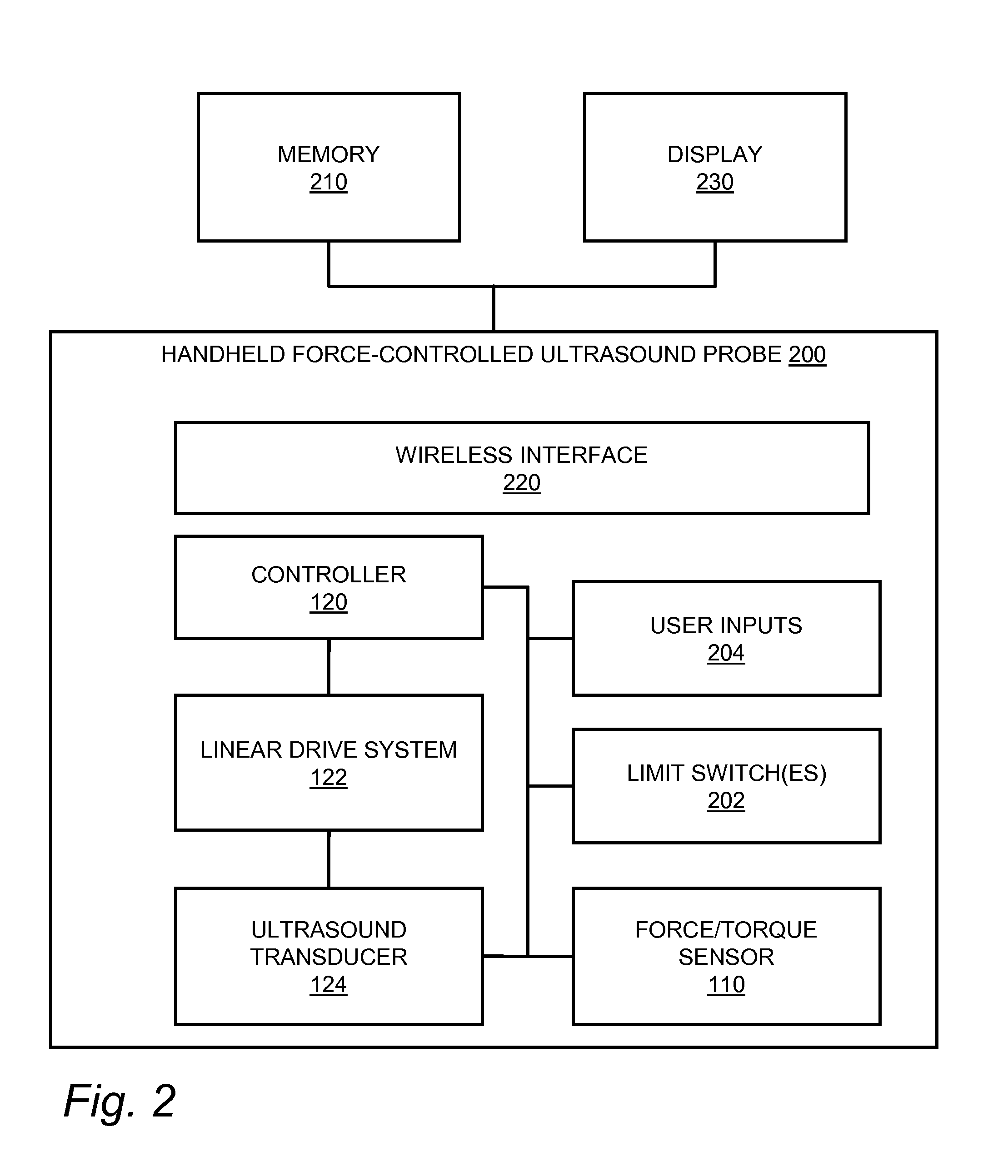
[0018]FIG. 2 is a schematic view of a handheld ultrasound probe.

[0019]FIG. 3 is a flowchart for a process for force-controlled acquisition of ultrasound images.

[0020]FIG. 4 depicts a lumped parameter model of the mechanical system of a probe as described herein.

[0021]FIG. 5 is a flowchart depicting operating modes of a force-controlled ultrasound probe.

DETAILED DESCRIPTION

[0022]The techniques described below allow real-time control of the contact force between an ultrasound probe and a target, such as a patient's body. This allows ultrasound technicians to take fixed- or variably-controlled-contact-force ultra...


PUM


Abstract

A handheld ultrasound probe includes force detection and feedback to permit control and/or characterization of applied forces. The probe may, for example, be operated to maintain a substantially constant applied force over a period of use independent of a user's hand position, or to measure applied forces that might contribute to deformation of an imaging subject.

Description

CROSS-REFERENCE TO RELATED APPLICATIONS[0001]This application is a continuation of U.S. patent application Ser. No. 12 / 972,461 filed Dec. 18, 2010, which claims the benefit of provisional application 61 / 287,886 filed Dec. 18, 2009 and entitled “HANDHELD FORCE-CONTROLLED ULTRASOUND PROBE,” each of which is hereby incorporated by reference in its entirety.BACKGROUND[0002]Medical imaging technologies permit viewing of internal body structures and anatomy without invasive surgical procedures. Ultrasound imaging, in particular, allows a physician to visualize internal details of soft tissue, organs, and the like by propagating sonic waves through a body and detecting sonic waves as they reflect off various internal structures. While current ultrasound techniques can provide useful diagnostic and treatment information, data for ultrasound imaging is generally captured with a handheld probe that is pressed against a body surface until suitable contact forces are achieved for imaging.[0003]...

Claims


Application Information

Patent Timeline
IPC IPC(8): A61B8/14
CPCA61B5/0048A61B5/6843A61B8/4209G01N29/32A61B8/4472G01N29/226G01N29/28A61B8/4281A61B5/0053
Inventor ANTHONY, BRIAN W.GILBERTSON, MATTHEW W.
Owner MASSACHUSETTS INST OF TECH
Why Eureka
Try Eureka