Image processing apparatus, image processing method, program, and program recording medium

Inactive Publication Date: 2011-09-01
CANON KK
View PDF17 Cites 25 Cited by
  • Summary
  • Abstract
  • Description
  • Claims
  • Application Information

AI Technical Summary

Benefits of technology

[0008]The present invention provides an image process

Problems solved by technology

During this time, the eye being examined (hereinafter this will be referred to as the subject's eye) may suddenly move or blink, resulting in a shift or distortion in the image.
However, such a shift or distortion in the image may not be recognized while the image is being captured.
Also, such a shift or distortion may be overlooked when the captured image data is checked after the image capturing is completed because of the vast amount of the image data.
Since this checking operation is not easy, the diagnosis workflow of a doctor is inefficient.
However, the known techniques have the following problems.
Therefore, although blinking can be detected, a shift or distortion in the image due to the movement of the subject's eye cannot be detected.
Thus, the image capturing state including the movement of the subject's eye cannot be obtained.
Therefore, when the eye greatly moves, the tomograms are corrected, but no accurate image can be generated.
Also, there is no concept to detect the image capturing state, which is the state of the subject's eye at the time the image is captured.

Method used


Image

  • Image processing apparatus, image processing method, program, and program recording medium
  • Image processing apparatus, image processing method, program, and program recording medium
  • Image processing apparatus, image processing method, program, and program recording medium

Examples


first embodiment

[0044]An image processing apparatus according to the present embodiment generates an integrated image from tomogram volume data when tomograms of a subject's eye (eye serving as an examination target) are obtained, and determines the accuracy of the captured images by using the continuity of image features obtained from the integrated image.

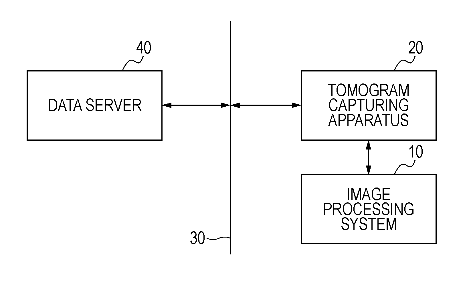
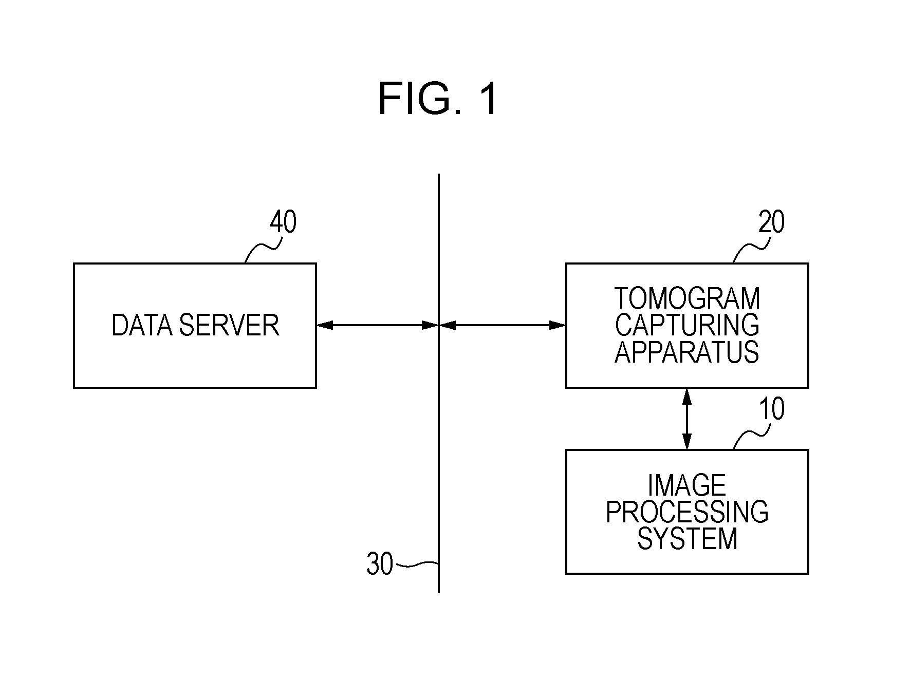
[0045]FIG. 1 is a block diagram of devices connected to an image processing system 10 according to the present embodiment. As illustrated in FIG. 1, the image processing system 10 is connected to a tomogram capturing apparatus 20 and a data server 40 via a local area network (LAN) 30 such as Ethernet (registered trademark). The connection with these devices may be established using an optical fiber or an interface such as universal serial bus (USB) or Institute of Electrical and Electronic Engineers (IEEE) 1394. The tomogram capturing apparatus 20 is connected to the data server 40 via the LAN 30 such as Ethernet (registered trademark). The conne...

second embodiment

[0090]In the present embodiment, the details of the process performed by the image processing unit 252 are different. A description of portions of the process that are the same as or similar to the first embodiment will be omitted.

[0091]The image processing unit 252 detects an edge region in the integrated image. By detecting an edge region parallel to the scanning direction at the time tomograms were captured, the image processing unit 252 obtains, in numeric terms, the degree of similarity between cross-sectional images constituting tomogram volume data.

[0092]When an integrated image is generated from tomogram volume data obtained by capturing tomograms of a position away from the retina since the eye moved at the time the tomograms were captured, the integrated value is different at a place where there is a positional shift due to the difference in the retina layer thickness.

[0093]Alternatively, when the eye blinked at the time the tomograms were captured, the integrated value be...

third embodiment

[0099]In the present embodiment, the image processing unit 252 performs a frequency analysis based on Fourier transform to extract frequency characteristics. The determining unit 253 determines whether items of tomogram volume data are continuous, in accordance with the strength in a frequency domain.

[0100]FIG. 10A is an illustration of an example of an integrated image. FIG. 10B is an illustration of an example of a power spectrum. Specifically, FIG. 10A illustrates an integrated image Pb generated when image capturing is unsuccessful due to a positional shift, and FIG. 10B illustrates a power spectrum Pb″ of the integrated image Pb. When there is a positional shift due to the eye movement at the image capturing time or when an eye blinks at the image capturing time, a spectrum orthogonal to the scanning direction at the time of image capturing using OCT is detected.

[0101]Using these results, the determining unit 253 determines the continuity of tomograms and the image capturing st...


PUM


Abstract

An image processing unit obtains information indicating continuity of tomograms of a subject's eye, and a determining unit determines the image capturing state of the subject's eye on the basis of the information obtained by the image processing unit.

Description

TECHNICAL FIELD [0001]The present invention relates to an image processing system that supports capturing of an image of an eye, and more particularly, to an image processing system using tomograms of an eye.BACKGROUND ART[0002]For the purpose of conducting early diagnoses of various diseases that occupy the top places of the causes of adult diseases and blindness, eye examinations are widely conducted. In examinations and the like, it is requested to find diseases of the entirety of an eye. Therefore, examinations using images of a wide area of an eye (hereinafter called wide images) are essential. Wide images are captured using, for example, a retinal camera or a scanning laser ophthalmoscope (SLO). In contrast, eye tomogram capturing apparatuses such as an optical coherence tomography (OCT) apparatus can observe the three-dimensional state of the interior of retina layers, and therefore, it is expected that these eye tomogram capturing apparatuses are useful in accurately conduct...

Claims


Application Information

Patent Timeline
IPC IPC(8): G06K9/00H04N7/18
CPCG06T7/0028A61B3/102G06T2207/30041G06T2207/10101G06T7/33A61B3/10G01N21/17A61B3/12G06T1/00
Inventor IWASE, YOSHIHIKOIMAMURA, HIROSHIFURUKAWA, DAISUKE
Owner CANON KK
Why Eureka
Try Eureka