High-resolution infrared imaging for enhanced detection, diagnosis, and treatment of cutaneous lesions

a high-resolution, infrared imaging technology, applied in the field of medical diagnosis, can solve the problems of unresolved quantitative response of healthy skin tissue or melanoma lesions, inability of systemic agents to significantly extend the lifespan of patients with advanced disease, and inability to quantify such response from healthy skin tissue or from melanoma lesions

Inactive Publication Date: 2011-09-22
THE JOHN HOPKINS UNIV SCHOOL OF MEDICINE
View PDF18 Cites 21 Cited by
  • Summary
  • Abstract
  • Description
  • Claims
  • Application Information

AI Technical Summary

Problems solved by technology

The reported 1-year survival rates for patients with advanced melanoma range from 40% to 60%, and systemic agents are not currently available to significantly extend the lifespan of patients with advanced disease.
In particular, describing the thermal response of chemically and metabolically active multilayered samples constitutes an important problem.
However, the problem of quantifying such response from healthy skin tissue or from melanoma lesions still remains unresolved.

Method used


Image

  • High-resolution infrared imaging for enhanced detection, diagnosis, and treatment of cutaneous lesions
  • High-resolution infrared imaging for enhanced detection, diagnosis, and treatment of cutaneous lesions
  • High-resolution infrared imaging for enhanced detection, diagnosis, and treatment of cutaneous lesions

Examples


Embodiment Construction

[0037]Some embodiments of the current invention are discussed in detail below. In describing embodiments, specific terminology is employed for the sake of clarity. However, the invention is not intended to be limited to the specific terminology so selected. A person skilled in the relevant art will recognize that other equivalent components can be employed and other methods developed without departing from the broad concepts of the current invention. All references cited herein are incorporated by reference as if each had been individually incorporated.

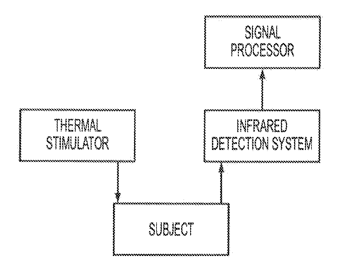
[0038]FIG. 1 is a schematic diagram of an embodiment of the invention. A thermal stimulator delivers a thermal stimulation to a subject under observation. The thermal stimulator can deliver a cooling stress by, for example, blowing cold air using a tube. Water, ice or a cold plate can also be used for the cooling stress. The thermal stimulator can deliver a heating stress by, for example, blowing warm air. Water or warm plate can also...


PUM


Abstract

A medical diagnosis system, comprising: a thermal stimulator; an infrared detection system constructed and arranged to detect infrared radiation from at least a portion of a subject under observation to provide an output signal from the portion of the subject after undergoing thermal stimulation from said thermal stimulator; and a signal processor in communication with the infrared detection system to receive the output signal from the infrared detection system, wherein the signal processor determines a measured thermal response of the portion of the subject to the thermal stimulation and compares the measured thermal response to an expected thermal response to determine a presence of an abnormality.

Description

CROSS-REFERENCE TO RELATED APPLICATIONS[0001]This application claims priority to U.S. Provisional Application No. 61 / 118,783 filed Dec. 1, 2008 and U.S. Provisional Application No. 61 / 122,770 filed Dec. 16, 2008, the entire contents of which are hereby incorporated by reference.[0002]The U.S. Government has a paid-up license in this invention and the right in limited circumstances to require the patent owner to license others on reasonable terms as provided for by the terms of Grant No.: 0651981, awarded by the National Science Foundation.BACKGROUND[0003]1. Field of Invention[0004]The current invention relates to medical diagnosis, and more particularly to medical diagnosis of lesions using infrared imaging.[0005]2. Discussion of Related Art[0006]Melanoma incidence is increasing at one of the fastest rates for all cancers in the United States with a current lifetime risk of 1 in 58. Over 60,000 patients are expected to be diagnosed with melanoma in the US with more than 8,000 deaths...

Claims


Application Information

Patent Timeline
Patent Type & Authority Applications(United States)
IPC IPC(8): A61F7/00
CPCA61B5/0059G01J2005/0077G01J5/0025A61B5/015
Inventor HERMAN, CILAALANI, RHODAPIRTINI CETINGUL, MUGE
Owner THE JOHN HOPKINS UNIV SCHOOL OF MEDICINE
Why Eureka
Try Eureka