
Medical diagnostic method and apparatus

a diagnostic method and a technology for medical diagnosis, applied in the field of medical diagnosis methods and equipment, can solve the problems of adding to the difficulty of establishing the perimeter, and achieve the effects of increasing the clinical value of the bladder volume determination, convenient and convenient use, and convenient and convenient carrying

Inactive Publication Date: 2011-10-27
SIGNOSTICS LTD
View PDF10 Cites 11 Cited by
  • Summary
  • Abstract
  • Description
  • Claims
  • Application Information

AI Technical Summary

Benefits of technology

"The patent describes a method for accurately determining the volume of bodily structures, such as the bladder, using ultrasound images. The method is particularly useful for clinical applications where the volume of urine in the bladder needs to be measured. The method involves acquiring multiple ultrasound images, estimating the location of the structure's perimeter, calculating the cross-sectional area, and combining the perimeter and area information to calculate the volume. The method takes into account the quality of the ultrasound images and the tissue response to ultrasound. The patent also describes a handheld ultrasound device that is easy to use, portable, and battery powered, with low power consumption. The technical effects of the patent are improved accuracy and ease of use for determining bodily structure volume, particularly for clinical applications."

Problems solved by technology

The varying quality of the ultrasound images, including image noise adds to the difficulty of establishing this perimeter.

Method used


Image

  • Medical diagnostic method and apparatus
  • Medical diagnostic method and apparatus
  • Medical diagnostic method and apparatus

Examples


Embodiment Construction

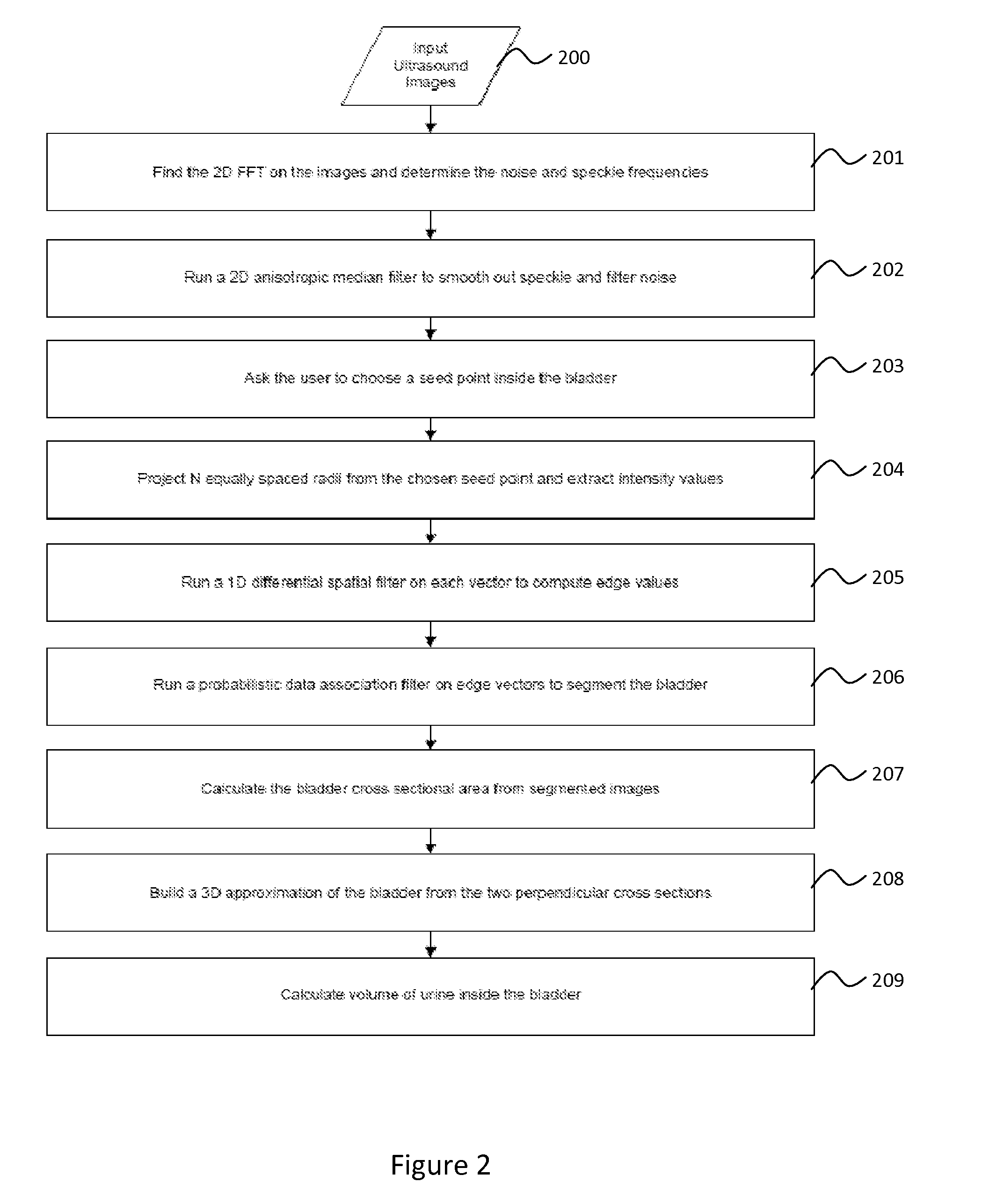
[0037]Now referring to the illustrations and in particular to FIG. 2, there is shown a flow chart of the method of the invention. The method illustrated is a method for determining the volume of a bladder. However, the method can be used for determining the volume of any organ or body structure which will show a reasonably distinct perimeter in an ultrasound scan. This may include the abdominal aorta, the prostate or other organs.

[0038]The first step 200 requires the input of ultrasound images. Preferably, these are two or more cross sectional images of the bladder, taken in directions substantially orthogonal to each other.

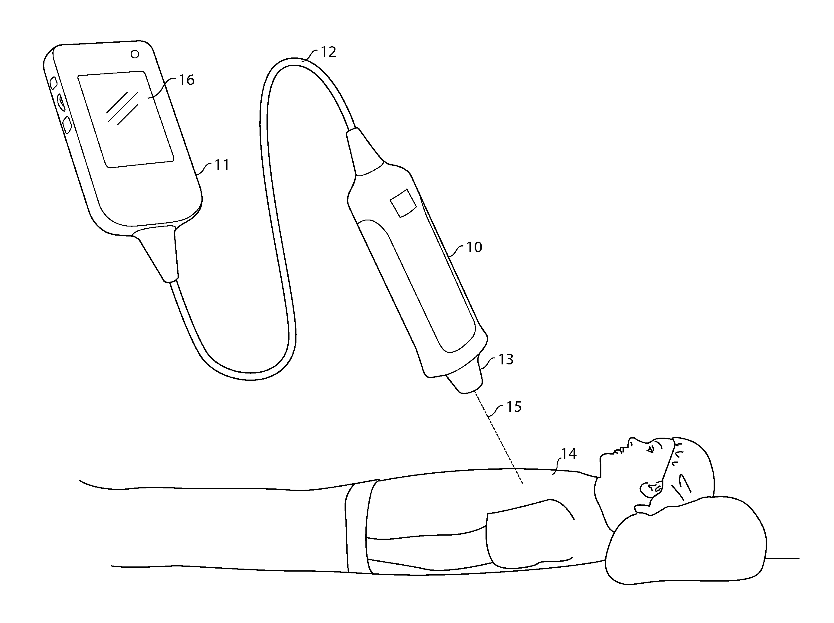
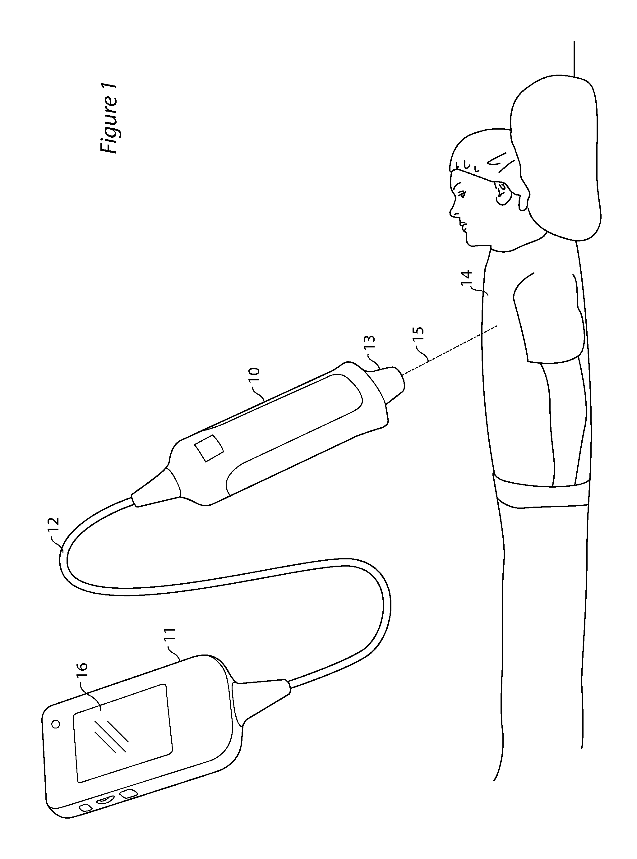
[0039]These images may be produced by any convenient means, however it is useful for these to be made by an inexpensive hand held ultrasound machine. This greatly expands the usefulness of the determination of bladder volume, since such a machine may available in contexts such as nursing home use, or visiting medical staff use where a full size machine cannot eco...


PUM


Abstract

A method and apparatus for determining the volume of a bodily structure including the steps of acquiring at least two ultrasound images, estimating the location of the perimeter of a structure of interest as viewed in cross section in each said ultrasound image, calculating the cross sectional area of such structure as so viewed, and combining the perimeter and area information from the at least two images to calculate the volume of the structure.

Description

TECHNICAL FIELD[0001]The present invention relates to a method and apparatus for determining the volume of an organ by use of ultrasound imaging. In particular the method and apparatus may be applicable to determination of the volume of a human or animal bladder.BACKGROUND ART[0002]At times it is clinically useful to know the area or spatial volume of a structure within the body. Examples are the cross section of the abdominal aorta and the volume of the bladder. Non-invasive bladder volume measurement techniques with ultrasound sonography have been described in the art. In principle, ultrasound scanning measures distance based on echo travel time. Early echo techniques used a single ultrasound transducer and echo presentation was recorded as echo amplitude versus depth. A method for determining bladder volume to determine residual urine volume based on distance measurement to the dorsal posterior bladder wall was described in the 1960's. The method was not adjusted to specific, fil...

Claims


Application Information

Patent Timeline
Patent Type & Authority Applications(United States)
IPC IPC(8): A61B8/00
CPCA61B8/14A61B8/145G06T2207/30084G06T2207/10132G06T7/602G06T7/62G06T2207/20182G06T2207/20192
Inventor EL-AKLOUK, ESSABRYDON, JOHN
Owner SIGNOSTICS LTD
Learn More