Glaucoma Treatment Device

a glaucoma and treatment device technology, applied in the field of glaucoma treatment devices, can solve the problems of optic nerve damage, optic nerve damage, abnormal high pressure, etc., and achieve the effects of reducing scarring, minimizing hypotonia, and eliminating complications such as endophthalmitis and leakag

Inactive Publication Date: 2011-12-15
NOVARTIS AG
View PDF7 Cites 87 Cited by
  • Summary
  • Abstract
  • Description
  • Claims
  • Application Information

AI Technical Summary

Benefits of technology

[0008]Disclosed are devices and methods for treatment of eye disease such as glaucoma. A shunt is placed in the eye wherein the shunt provides a fluid pathway for the flow or drainage of aqueous humour from the anterior chamber to the suprachoroidal space. The shunt is implanted in the eye using a delivery system that uses a minimally invasive procedure, as described below. By guiding fluid directly into the supraciliary or suprachoroidal space rather than to the surface of the eye, complications commonly encountered with conventional glaucoma surgery should be avoided. Shunting aqueous fluid flow directly into the supraciliary or suprachoroidal space should minimize scarring since the angle region is populated with a single line of non-proliferating trabecular cells. Shunting aqueous flow directly into the supraciliary or suprachoroidal space should minimize hypotony and also potentially eliminate complications such as endophthalmitis and leaks since an external filtering bleb is not the goal of surgery. The device described herein is designed to enhance aqueous flow through the normal outflow system of the eye with minimal to no complications. Any of the procedures and device described herein can be performed in conjunction with other therapeutic procedures, such as laser iridotomy, laser iridoplasty, and goniosynechialysis (a cyclodialysis procedure).

Problems solved by technology

It is known that glaucoma results in abnormally high pressure in the eye, which leads to optic nerve damage.
Over time, the increased pressure can cause damage to the optic nerve, which can lead to blindness.
The large market is mostly due to the fact that there are not any effective surgical alternatives that are long lasting and complication-free.
Unfortunately, drug treatments need much improvement, as they can cause adverse side effects and often fail to adequately control intraocular pressure.
Moreover, patients are often lackadaisical in following proper drug treatment regimens, resulting in a lack of compliance and further symptom progression. treatment regimens, resulting in a lack of compliance and further symptom progression.
Such a procedure can be quite traumatic as the implants are large and can result in various adverse events such as infections and scarring, leading to the need to re-operate.

Method used


Image

  • Glaucoma Treatment Device
  • Glaucoma Treatment Device
  • Glaucoma Treatment Device

Examples


Embodiment Construction

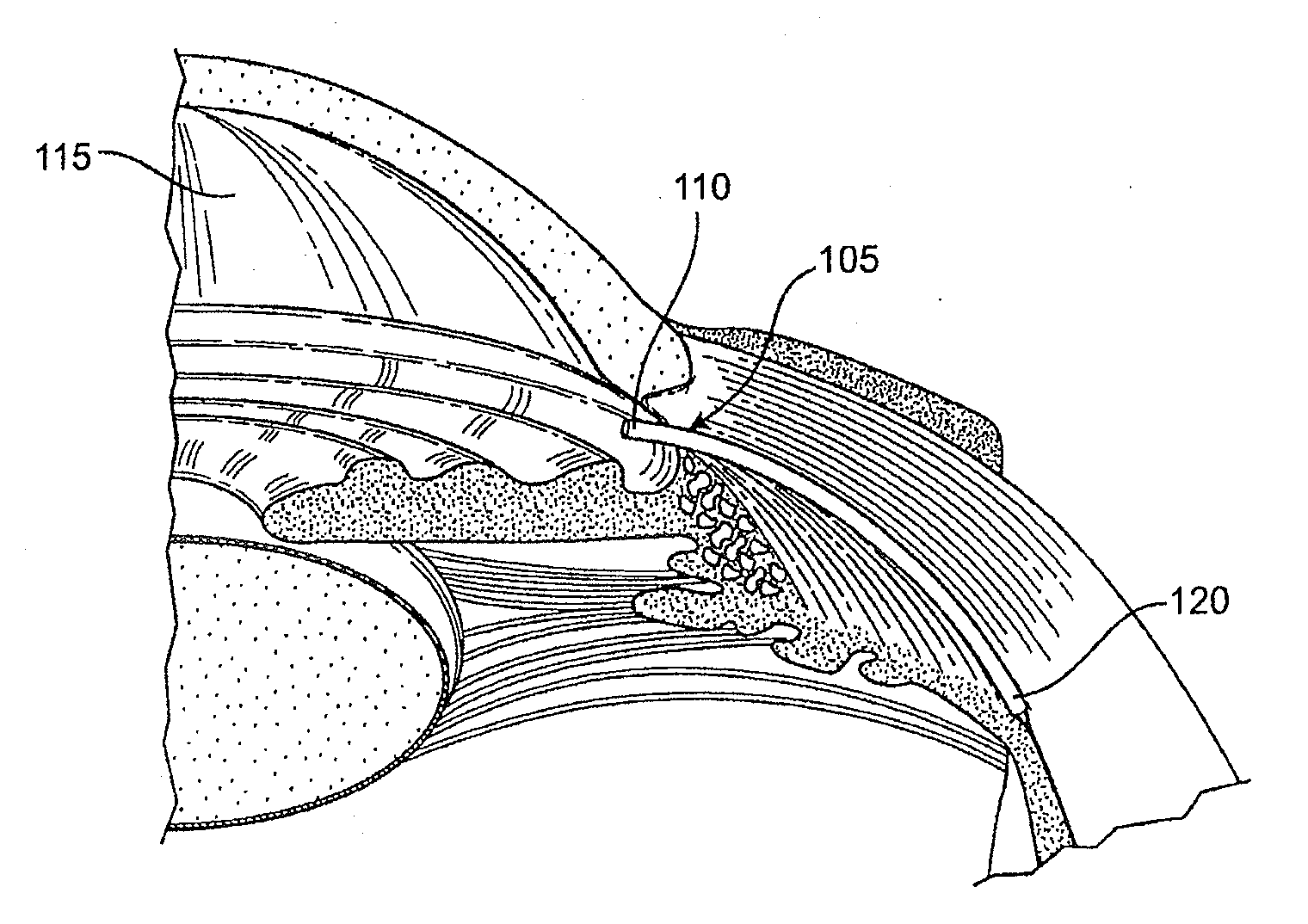
[0085]FIG. 1 is a cross-sectional, perspective view of a portion of the eye showing the anterior and posterior chambers of the eye. A shunt 105 is positioned inside the eye such that a proximal end 110 is located in the anterior chamber 115 and a distal end 120 is located in the suprachoroidal space (sometimes referred to as the perichoroidal space). The shunt 105 is illustrated in FIG. 1 as an elongate element having one or more internal lumens through which aqueous humour can flow from the anterior chamber 115 into the suprachoroidal space. Embodiments of the shunt 105 with various structural configurations are described in detail below.

Exemplary Eye Anatomy

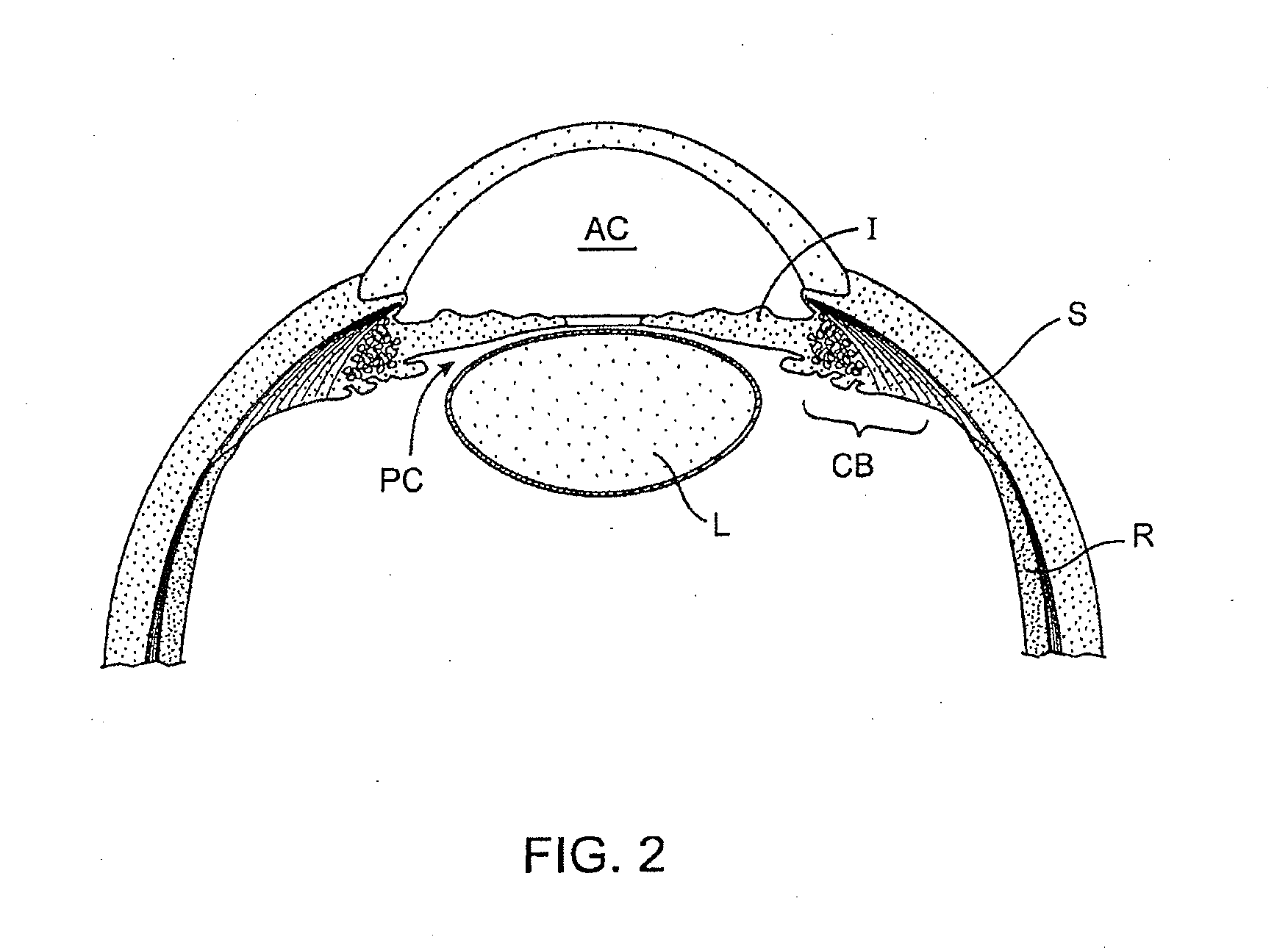
[0086]FIG. 2 is a cross-sectional view of a human eye. The eye is generally spherical and is covered on the outside by the sclera S. The retina R lines the inside posterior half of the eye. The retina registers the light and sends signals to the brain via the optic nerve. The bulk of the eye is filled and supported by the vitre...


PUM


Abstract

Methods and devices are adapted for implanting into the eye. An incision is formed in the cornea of the eye and a shunt is inserted through the incision into the anterior chamber of the eye. The shunt includes a fluid passageway. The shunt is passed along a pathway from the anterior chamber through the scleral spur of the eye into the suprachoroidal space and positioned in a first position such that a first portion of the fluid passageway communicates with the anterior chamber and a second portion of the fluid passageway communicates with the suprachoroidal space to provide a fluid passageway between the suprachoroidal space and the anterior chamber.

Description

RELATED APPLICATIONS[0001]This application is a continuation of co-pending U.S. patent application Ser. No. 11 / 615,810, filed Dec. 22, 2006, entitled “GLAUCOMA TREATMENT DEVICE,” which claims the benefit of priority under 35 U.S.C. §119(e) of U.S. Provisional Application Ser. Nos. 60 / 759,835, filed Jan. 17, 2006; 60 / 783,632, filed Mar. 17, 2006; and 60 / 824,396, filed Sep. 1, 2006, each of which are entitled “GLAUCOMA TREATMENT DEVICE.”[0002]The subject matter of each of the above-noted applications is incorporated by reference in its entirety by reference thereto.BACKGROUND[0003]This disclosure relates generally to methods and devices for use in treating glaucoma. The mechanisms that cause glaucoma are not completely known. It is known that glaucoma results in abnormally high pressure in the eye, which leads to optic nerve damage. Over time, the increased pressure can cause damage to the optic nerve, which can lead to blindness. Treatment strategies have focused on keeping the intra...

Claims


Application Information

Patent Timeline
Patent Type & Authority Applications(United States)
IPC IPC(8): A61F9/00A61F2/04
CPCA61K9/0051A61F2/142A61F2/14A61F9/0017A61M2202/04A61B19/22A61F9/0133A61F11/00A61M27/002A61F9/0008A61F9/00781A61M2210/0612A61F9/007A61B19/00A61F9/00736A61F2250/0001A61F2250/0096A61F2250/0097A61B34/70A61B90/00
Inventor DE JUAN, JR., EUGENEBOYD, STEPHENDEEM, MARK E.GIFFORD, III, HANSON S.ROSENMAN, DAN
Owner NOVARTIS AG
Why Eureka
Try Eureka