Eureka AIR delivers breakthrough ideas for toughest innovation challenges, trusted by R&D personnel around the world.

Full body plethysmographic chamber incorporating photoplethysmographic sensor for use with small non-anesthetized animals

a full-body plethysmographic and sensor technology, applied in the field of photoplethysmographic readings for animal research, can solve the problems of inability to obtain additional parameters, difficulty in obtaining accurate global figures for animal testing, and difficulty in obtaining techniques in humans, and achieves significant blood flow, easy affixed attachment mechanism, and inherent bite resistance to the sensor platform

Inactive Publication Date: 2011-12-22
STARR LIFE SCI
View PDF5 Cites 10 Cited by
  • Summary
  • Abstract
  • Description
  • Claims
  • Application Information

AI Technical Summary

Benefits of technology

[0038]A noninvasive photoplethysmographic sensor for mobile animals such as small rodents, namely rats and mice in a full body plethysmographic chamber is useful such as in a laboratory research environment. The noninvasive photoplethysmographic sensor may be a neck clip or collar which provides an easily affixed attachment mechanism. The neck location will provide significant blood flow under all conditions and also offers inherent bite resistance to the sensor platform. The system of the present invention provides a commutator for the sensor wires for allowing untwisting of the system wires.

Problems solved by technology

Accurate global figures for animal testing are difficult to obtain, or to verify.
The technique in humans requires moderately complex coaching and instruction for the subject.
The limitation in the existing systems is that they have not allowed for additional parameters to be obtained from the animal in the chamber.
The concept is similar to today's conventional pulse oximetry but suffered due to unstable photocells and light sources and the method was not used clinically.
This use was limited to pulmonary functions.
However, consideration must be made for the particular subject or range of subjects in the design of the pulse oximeter, for example the sensor must fit the desired subject (e.g., a medical pulse oximeter for an adult human finger simply will not adequately fit onto a mouse finger or paw; and regarding signal processing the signal areas that are merely noise in a human application can represent signals of interest in animal applications due to the subject physiology).
This approach has proven impractical as the human based systems can only stretch so far and this approach has limited the use of such adapted oximeters.
For example, these adapted human oximeters for animals have an upper limit of heart range of around 400 or 450 beats per minute which is insufficient to address mice that have a conventional heart rate of 400-800 beats per minute.
Expandable collars should not be used unless they are well tested, as poorly designed collars can be very problematic.
As a result, there is always the possibility of transmitter loss, icing up in winter, or of the collar becoming snagged by branches or even the animal's own legs.
For example, a breakaway bird body harness could easily impair wing movement as it is lost and result in mortality.
Radio and timer-controlled breakaways may be jammed by freezing or dirt, and also add to the size, weight and complexity of the transmitter package.
Paint or non-metallic reflective materials may be sewn or glued to collars and harnesses; however, this is likely not appropriate for cryptic species.
Adhesive tapes should also not be used as they are not very durable and may foul fur or feathers.
Size or weight limitations and the data precision required will also affect transmitter type and placement.
It is interesting to note that for pulse oximetry in cattle, off the shelf human oximeters were insufficient and a custom design was required.
None of the above solutions adequately address laboratory animal research applications using mobile animals in a full body plethysmographic chamber and more particularly, the prior art fails to adequately provide an efficient and noninvasive photoplethysmographic sensors for mobile animals such as small rodents, namely rats and mice in a full body plethysmographic chamber.

Method used


Image

  • Full body plethysmographic chamber incorporating photoplethysmographic sensor for use with small non-anesthetized animals
  • Full body plethysmographic chamber incorporating photoplethysmographic sensor for use with small non-anesthetized animals

Examples


Embodiment Construction

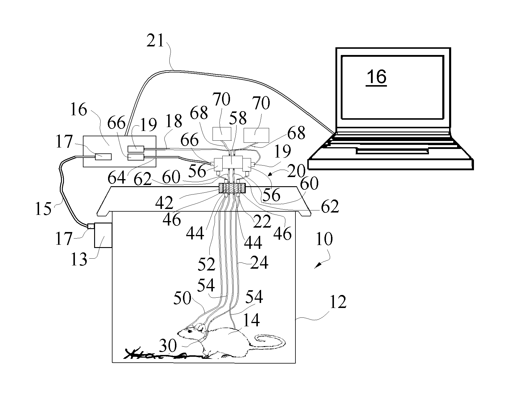
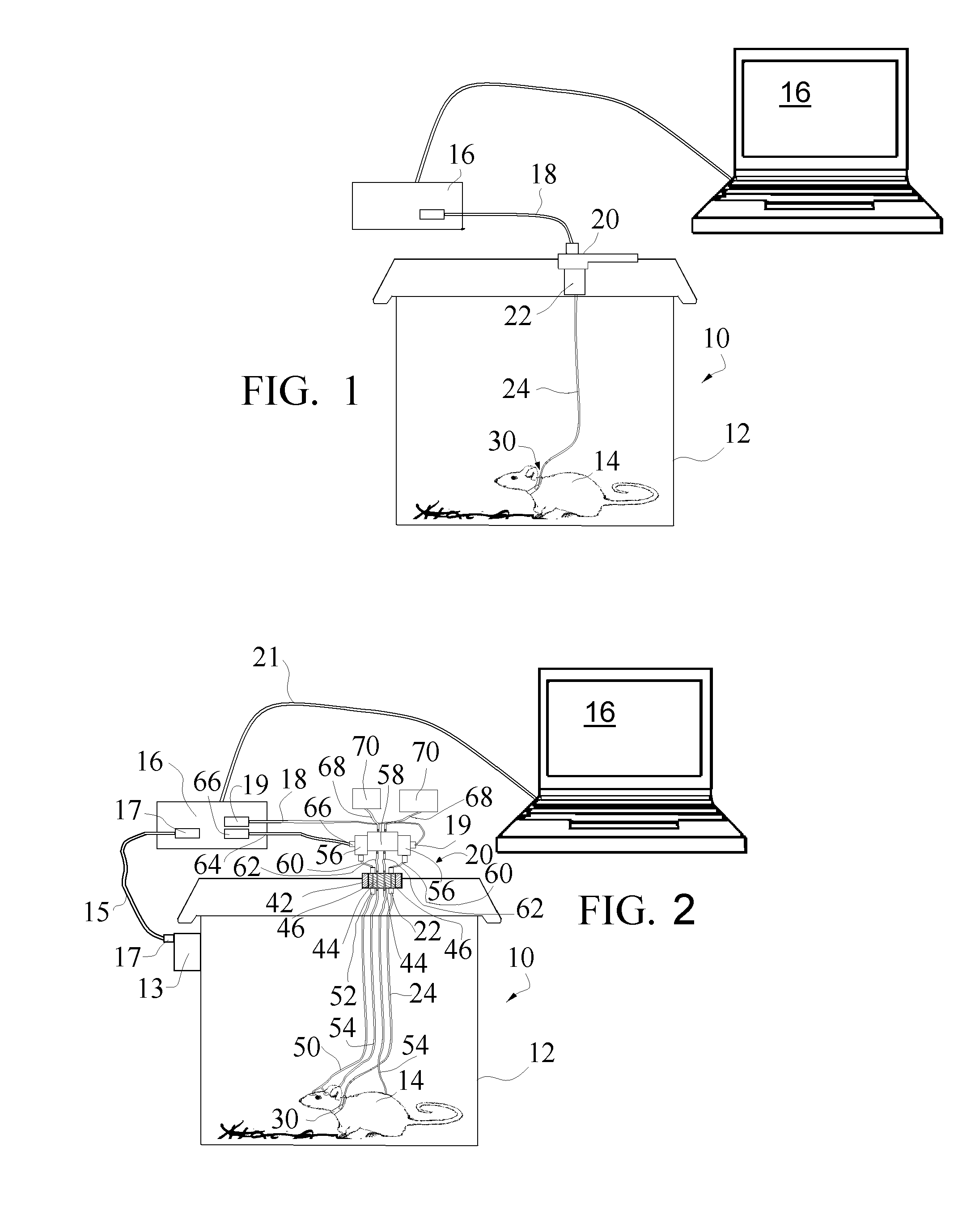
[0042]In summary, the present invention relates to a noninvasive plethysmographic chamber system 10 incorporating a photoplethysmographic sensor 30 for mobile animals, such as rats and mice 14 that are utilized in a sealed full body plethysmographic chamber 12. Photoplethysmographic measurements on laboratory animals have most often been accomplished on restrained and / or anesthetized animals. This limits the research that can be conducted. Further, in the pulse oximetry field there has been a lack of adequate photoplethysmographic sensors for small mice (and even small rats), until the advent of the Mouse Ox™ brand pulse oximeters by Starr Life Sciences. Prior to this development, commercially available pulse oximeters could provide heart rate data up to about 350 or 450 beats per minute (and even this range required special software modifications for some sensors), which were basically suitable for rats but not small mice given that the small mouse will have heart rates in the rang...


PUM


Abstract

A full body plethysmographic chamber includes a noninvasive photoplethysmographic sensor within the chamber. The noninvasive photoplethysmographic sensor is for mobile animals such as small rodents, namely rats and mice in the full body plethysmographic chamber and is useful in a laboratory research environment. The noninvasive photoplethysmographic sensor may be a neck clip or collar which provides an easily affixed attachment mechanism. The neck location will provide significant blood flow under all conditions and also offers inherent bite resistance to the sensor platform. The system of the present invention provides a commutator for the sensor wires for allowing untwisting of the system wires.

Description

[0001]The present invention claims priority of U.S. Provisional Patent Application Ser. No. 61 / 286,368 entitled “Photoplethysmographic Sensor for use in Full Body Plethysmographic Chamber on Small Non-Anesthetized Animals” filed Dec. 14, 2009.BACKGROUND OF THE INVENTION[0002]1. Field of the Invention[0003]The present invention relates to photoplethysmographic readings for animal research and more particularly, the present invention is directed to a full body plethysmographic chamber incorporating a noninvasive photoplethysmographic sensor for mobile animals such as small rodents.[0004]2. Background Information[0005]In general, medical research begins on animals, and among the animals used in research, teaching, and testing, mice and rats comprise, by far, the largest majority of all experimental mammals. Accurate global figures for animal testing are difficult to obtain, or to verify. The Nuffield Council on Bioethics reports that global annual estimates range from 50 to 100 million...

Claims


Application Information

Patent Timeline
Patent Type & Authority Applications(United States)
IPC IPC(8): A61B5/1455
CPCA61B5/0806A61B5/02416
Inventor HETE, BERNARD F.
Owner STARR LIFE SCI
Why Eureka
Eureka Blog
Learn More