Detection, measurement, and imaging of cells such as cancer and other biologic substances using targeted nanoparticles and magnetic properties thereof

a technology of targeted nanoparticles and cancer cells, applied in the field of in vivo detection and measurement of cells or substances using targeted nanoparticles, can solve the problems of low positive diagnosis rate, high false diagnosis rate, and no 100% accurate detection

Active Publication Date: 2012-02-09
IMAGION BIOSYST INC
View PDF5 Cites 17 Cited by
  • Summary
  • Abstract
  • Description
  • Claims
  • Application Information

AI Technical Summary

Benefits of technology

[0026]In some embodiments of the present invention, the size and extent of a tumor can be determined by computer-aided analysis using techniques such as minimum norm and multipole expansions. In some embodiments of the present invention, the location of the bound nanoparticles can be presented in combination with image information such as can be obtained from Xray instruments, photographs, or MRIs. In some embodiments of the present invention, measurements on phantoms can be used in combination with electromagnetic theory to determine location accuracy and detection sensitivity at various depths. In some embodiments of the present invention, the magnetic moment measured can be used to determine a number of nanoparticles bound to targeted cells, or a number of targeted cells having bound nanoparticles, using the characteristic that the magnetic moment is linearly related to the number of bound nanoparticles.
[0027]The present invention is described in terms of various example applications, including applications to the measurement of specific types of cancer cells. The invention is not limited to such examples, however. The invention can be useful in determining properties such as the presence of a biological substance in a sample, the location of a quantity of a biological substance in a sample, the quantity of a biological substance in a sample (e.g., the number of cancer cells in a tumor), or a combination thereof.

Problems solved by technology

Many common methods used for clinical purposes for detection of cancer are non-specific, i.e., they cannot distinguish between cancerous or benign tumors and none lead to 100% accurate detection.
The available methods all have disadvantages and weaknesses, resulting in high rates of false diagnosis and too low a rate of positive diagnosis, together leading to increased mortality rates.
Artifacts can be caused by healthy tissue and give rise to false positive results.
Irregularities in tissue such as scarring can cause non-uniform scattering of the x-rays rendering mammograms ineffective.
Overall, the number of false positives in x-ray imaging of cancer remains high and the x-ray method cannot detect early-stage tumors.
Ultrasound has excellent contrast resolution but suffers from diminished spatial resolution compared to x-rays and other imaging techniques.
MRI is used to follow up on potential problem areas seen during x-ray scans; however, the expense of a MRI scan often prohibits its use.
Dynamic Contrast Enhanced (DCE) MRI potentially distinguishes between benign and cancerous tumors but produces a number of false positives.
The expense of MRI limits its application as a screening tool.
Existing imaging methods often do not identify lesions until significant growth has occurred.
At present, none of these methods have specificity regarding tumor type using differences in tissue properties between cancerous and non-cancerous tissue.

Method used


Image

  • Detection, measurement, and imaging of cells such as cancer and other biologic substances using targeted nanoparticles and magnetic properties thereof
  • Detection, measurement, and imaging of cells such as cancer and other biologic substances using targeted nanoparticles and magnetic properties thereof
  • Detection, measurement, and imaging of cells such as cancer and other biologic substances using targeted nanoparticles and magnetic properties thereof

Examples


Embodiment Construction

[0058]The present invention is described in the context of various example embodiments and applications. In some of the description, the term “detection” is used for brevity; the invention can provide for the detection of the presence of cells or substances, measurement of the number of cells or amount of substance, determination of the location of cells or substance, determination of the change or rate of change in the preceding, and similar determinations, all of which are included in the term “detecting.”

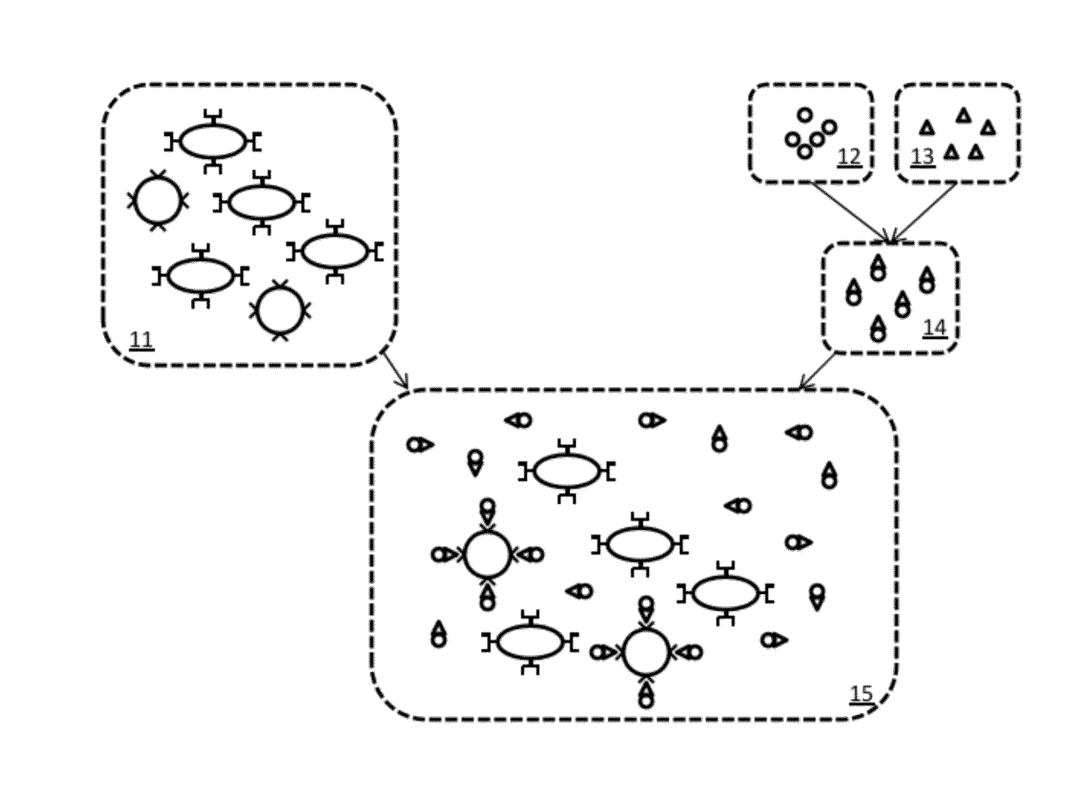
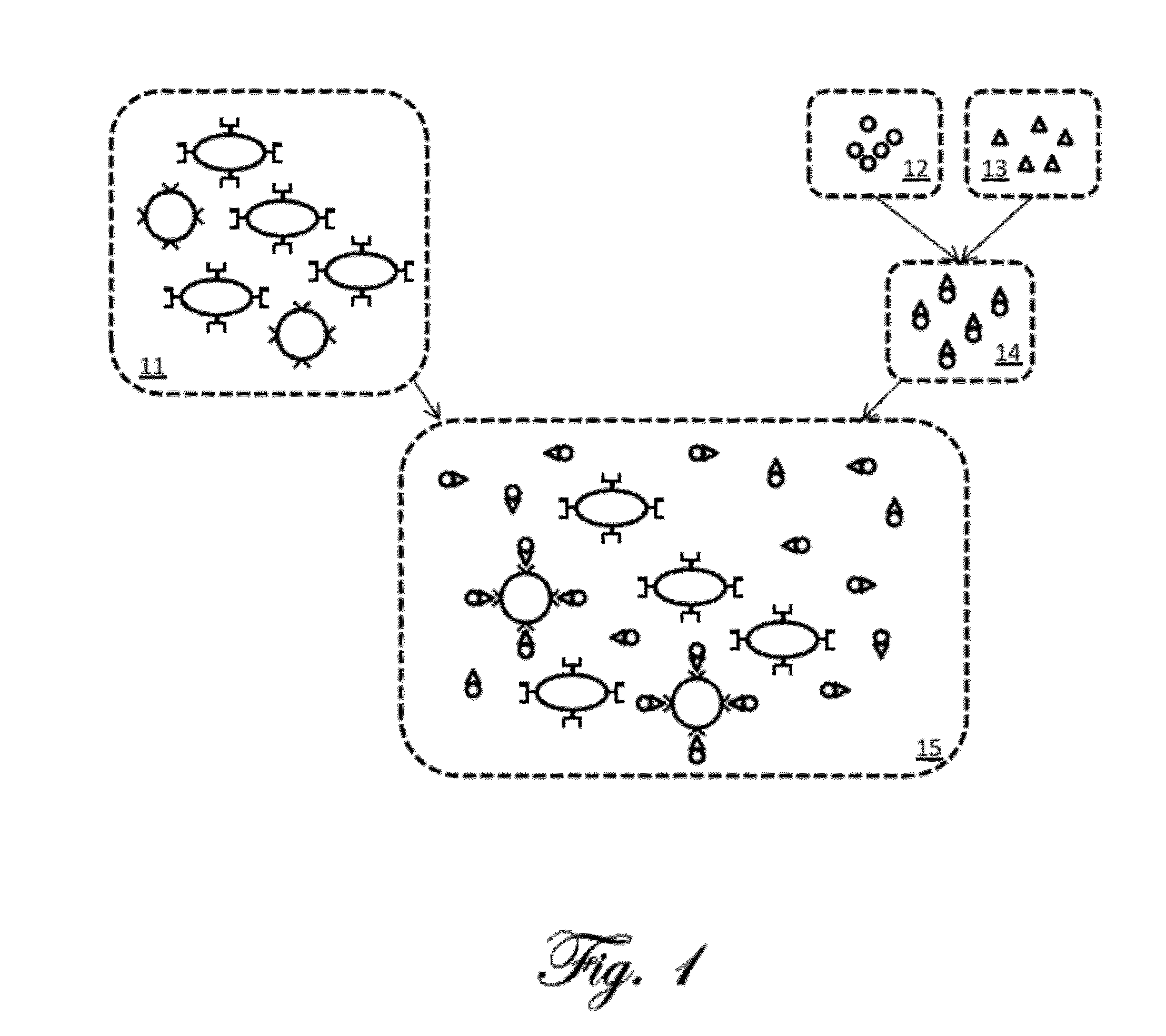
[0059]A simplified example of magnetic relaxation measurement according to the present invention is first described. FIG. 1 is a schematic illustration of an example preparation of a sample for measurement according to the present invention. The illustrations in the figure are highly simplified and intended for ease of explanation only, and are not intended to represent the actual shapes, sizes, proportions, or complexities of the actual materials involved. A portion of the tissu...


PUM


Abstract

The present invention can provide a method of determining the presence, location, quantity, or a combination thereof, of a biological substance, comprising: (a) exposing a sample to a plurality of targeted nanoparticles, where each targeted nanoparticle comprises a paramagnetic nanoparticle conjugated with one or more targeting agents that preferentially bind with the biological substance, under conditions that facilitate binding of the targeting agent to at least one of the one or more biological substances; (b) subjecting the sample to a magnetic field of sufficient strength to induce magnetization of the nanoparticles; (c) measuring a magnetic field of the sample after decreasing the magnetic field applied in step b below a threshold; (d) determining the presence, location, quantity, or a combination thereof, of the one or more biologic substances from the magnetic field measured in step (c).

Description

CROSS REFERENCE TO RELATED APPLICATIONS[0001]This invention claims priority as a continuation in part of U.S. application Ser. No. 11 / 940,673 filed Nov. 15, 2007, which application claimed priority to U.S. application 60 / 866,095;[0002]and as a continuation in part of U.S. application Ser. No. 12 / 337,554 filed Dec. 17, 2008, which application was a continuation in part of U.S. application Ser. No. 11 / 957,988 filed Dec. 17, 2007, which application was a continuation in part of U.S. application Ser. No. 11 / 940,673 filed Nov. 15, 2007;[0003]and as a continuation in part of PCT application PCT / US2010 / 051417 filed Oct. 5, 2010, which application claimed priority to U.S. application 61 / 248,775 filed Oct. 5, 2009, U.S. application 61 / 329,076 filed Apr. 28, 2010, and U.S. application 61 / 377,854 filed Aug. 27, 2010;[0004]and as a continuation in part of PCT application PCT / US2010 / 055729 filed Nov. 5, 2010, which application claimed priority to U.S. application 61 / 314,392 filed Mar. 16, 2010, ...

Claims


Application Information

Patent Timeline
Patent Type & Authority Applications(United States)
IPC IPC(8): A61B5/05
CPCA61B5/05A61B5/4088A61B5/415A61B5/416A61B5/418G01R33/0354G01R33/1269G01R33/1276
Inventor FLYNN, EDWARD R.
Owner IMAGION BIOSYST INC
Why Eureka
Try Eureka