Composition for stimulating bone growth and differentiation and method for isolating same

a technology of differentiation and differentiation, which is applied in the field of isolated bone, can solve the problems of poor clinical efficacy, poor clinical efficacy, and inability to achieve the effect of enhancing bone cell growth and particularly useful properties

Inactive Publication Date: 2005-12-29
THE UNIV OF QUEENSLAND
View PDF1 Cites 25 Cited by
  • Summary
  • Abstract
  • Description
  • Claims
  • Application Information

AI Technical Summary

Benefits of technology

[0011] However, contrary to WO 93 / 19096, the present inventors have surprising found that HS obtained from a specific tissue source may have particularly useful properties, in particular as a potential therapeutic and pharmaceutical composition. Although HS has been previously extracted from skin, brain, liver and cultured cells, HS has never been extracted from bone or bone precursor cells prior to this invention. The inventors were surprised to find that HS isolated from bone cells when applied to cells showed a greater increase in bone cell growth when compared with other sources of HS, as is described in more detail hereinafter.

Problems solved by technology

Although there have been advances in the area of implants anchored in bone tissue, current orthopaedic practices for repair of load-bearing bone are rather crude and often fail.
WO 93 / 19096 states that in contrast to the useful properties of oligosaccharides as therapeutics, HS is not particularly useful as a therapeutic.
In fact, even fragments of HS (i.e. oligosaccharides prepared from enzyme digested HS), are also not considered suitable for use as a therapeutic due to a resulting complex mixture of various molecular species having a wide range of different compositions and sizes.

Method used


Image

  • Composition for stimulating bone growth and differentiation and method for isolating same
  • Composition for stimulating bone growth and differentiation and method for isolating same
  • Composition for stimulating bone growth and differentiation and method for isolating same

Examples


example 1

Cell Culture and Radiolabelling

[0177] Bone precursor MC3T3 cells were grown in 250 ml tissue culture flasks in 5% FCS / DMEM in a 10% CO2 / air-humidified incubator. When isolating logarithmic growth HS, radiolabel was added 24 h post-passaging and the cells allowed to grow unhindered for 3 days. To isolate HS from contact-inhibited cells, media on the cells was changed to 0.5% FCS / DMEM post-confluence and radiolabelled (20 μCi / ml) 24 h after the media was changed. Cells were maintained at confluence for 3 days and then the media collected and frozen at −20° C. until required. Cell membranes were prepared in lysis buffer (1% Triton X100, 150 mM NaCl, 10 mM Tris pH 7.4, 2 mM EDTA, 0.5% NP 40, 0.1% SDS containing the protease inhibitors 1 mM sodium orthovanadate, 10 μg / ml leupeptin, 1 μg / ml aprotinin and 1 mM PMSF). The cellular ECM was collected with lysis buffer plus 6 M Urea.

example 2

Determination of metabolic Activity using WST-1

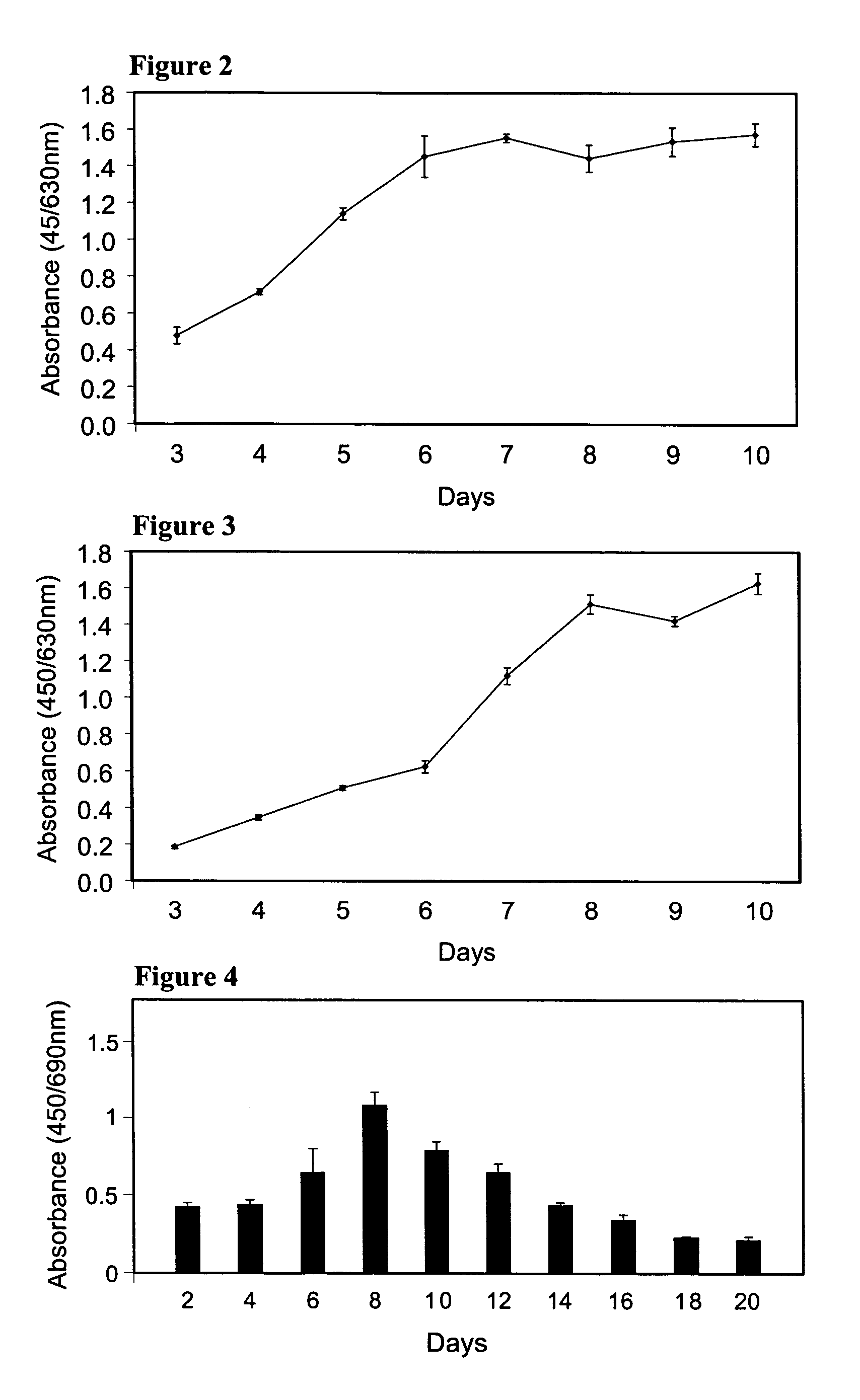
[0178] Unless otherwise indicated, MC3T3-E1 cells were plated at 5000 cells / cm2 into wells of a 96 well plate in triplicate, allocating 3 wells to each time point, and grown in osteogenic media for 3-10 days. The Cell Proliferation Reagent WST-1 (Roche Diagnostics, Singapore) was added to triplicate wells at each time point, diluted 1:10 into the media. The reaction was catalysed by the conversion of WST-1, a tetrazolium salt, into formazon by mitochondrial dehydrogenase, which directly correlates to the number of metabolically-active cells in the culture. The reaction is incubated for 37° C. for 30 min, liberating a red colour, and read at 450 nm with a reference wavelength of 630 nm on a Victor3™ Multilevel Plate Reader (Perkin Elmer, Boston, Mass., USA). A blank well containing only media was used for background correction due to discolouration by the media.

[0179] As the assay can be performed and read under sterile conditions, cel...

example 3

Determination of Cell Proliferation using BrdU

[0180] Cell proliferation was analysed with a Cell Proliferation ELISA colorimetric kit (Roche, Switzerland). MC3T3-E1 cells were incubated with 10 μM BrdU for 2 h at 37° C., denatured, fixed and incubated with anti-BrdU-POD for 90 min at RTP according to the manufacturer's instructions. The reaction was catalysed by the addition of a tetramethylbenzidine substrate solution and terminated after 15 min with 1 M H2SO4. The absorbance was read at 450 nm (with a reference of 690 nm) using a Bio-Rad® Benchmark™ Microplate Reader (Bio-Rad, Calif., USA) and corrected using blank and background controls.


PUM

PropertyMeasurementUnit
wavelengthaaaaaaaaaa
pHaaaaaaaaaa

Abstract

This invention relates to isolated heparan sulphate and use thereof to stimulate bone cell growth and differentiation. The invention also relates to use of heparan sulphate with implants, prosthesis and bioscaffolds to repair and regenerate bone. Such use may be for repair of damaged tissue including bone tissue, for example damage resulting from injury or defect.

Description

[0001] This application claims priority of Australian Provisional Application Ser. No 2004902408, filed May 7, 2004. FIELD OF THE INVENTION [0002] This invention relates to isolated heparan sulphate and use thereof to stimulate bone cell growth and differentiation. The invention also relates to use of heparan sulphate with implants, prosthesis and bioscaffolds to repair and regenerate bone. Such use may be for repair of damaged tissue including bone tissue, for example damage resulting from injury or defect. BACKGROUND OF THE INVENTION [0003] Although there have been advances in the area of implants anchored in bone tissue, current orthopaedic practices for repair of load-bearing bone are rather crude and often fail. Use of titanium-based materials has improved implants; however, there is still a need for methods and compositions that may assist or stimulate regeneration of natural bone for use with, or independently from, an implant. [0004] Humans and other animals are complex mult...

Claims


Application Information

Patent Timeline
Patent Type & Authority Applications(United States)
IPC IPC(8): A61K31/727A61K31/737A61P19/00C08B37/10
CPCA61K31/727C08B37/0078C08B37/0075A61K31/737A61P19/00
Inventor NURCOMBE, VICTORCOOL, SIMON
Owner THE UNIV OF QUEENSLAND
Why Eureka
Try Eureka