Endoscopic polarized multispectral light scattering scanning method

a scanning method and multi-spectral light technology, applied in the field of endoscopic polarized multi-spectral light scattering scanning method, can solve the problems of increasing the chance of successful treatment, poor diagnosis of adenocarcinoma, and affecting the treatment effect, so as to reduce the time delay

Active Publication Date: 2012-02-16
BETH ISRAEL DEACONESS MEDICAL CENT INC
View PDF12 Cites 22 Cited by
  • Summary
  • Abstract
  • Description
  • Claims
  • Application Information

AI Technical Summary

Benefits of technology

[0008]Embodiments of the invention may enable fast and reliable spectroscopic detection and diagnosis of dysplasia in patients with BE during endoscopy. Exemplary embodiments may provide an instrument and method for safely, quickly, and reliably surveying the entire length of Barrett's esophagus for endoscopically invisible dysplasia, thereby avoiding harms and risks associated with random biopsy surveillance.
[0013]According to another aspect of the invention, a method and system for obtaining diagnostic information from polarized LSS data of tissue is provided. The described method may be used to provide diagnostic infotmation corresponding to separate tissue locations, corresponding to tissue located along a line and / or corresponding to tissue over a two-dimensional area, as the invention is not limited in this respect. The described system may be used to analyze spectral data obtained from one or more tissue locations in vivo or from tissue samples ex vivo to produce data indicating a diagnostic condition at each tissue location. The described system may be used to obtain a real-time map of a diagnostic condition over an area of tissue during an endoscopy. Thus, an entire surface of the organ may be rapidly scanned and results of the scanning are analyzed with a reduced time delay. The system may also include an imaging system for providing an image of the tissue during an endoscopy. The system may enable biopsies at locations where a certain tissue condition has been identified or is suspected based on a map of nuclear tissue structure and / or a map of a diagnostic condition obtained by LSS. A map of nuclear tissue structure and / or a map of a diagnostic condition may be displayed along with an image of the same location during an endoscopy.

Problems solved by technology

Although the prognosis of patients diagnosed with adenocarcinoma may be poor, the chances of successful treatment increase significantly if the disease is detected at the dysplastic stage.
The surveillance of patients with BE for dysplasia may be challenging in at least three respects.
First, dysplasia may not be visible during routine endoscopy.
Second, the histopathologic diagnosis of dysplasia is problematic because there is poor interobserver agreement on the classification of a particular specimen, even among expert gastrointestinal pathologists.
Third, reliance on the histopathologic examination imposes a time delay between endoscopy and diagnosis, which may severely limit the diagnostic accuracy of the endoscopic procedure.

Method used


Image

  • Endoscopic polarized multispectral light scattering scanning method
  • Endoscopic polarized multispectral light scattering scanning method
  • Endoscopic polarized multispectral light scattering scanning method

Examples


Embodiment Construction

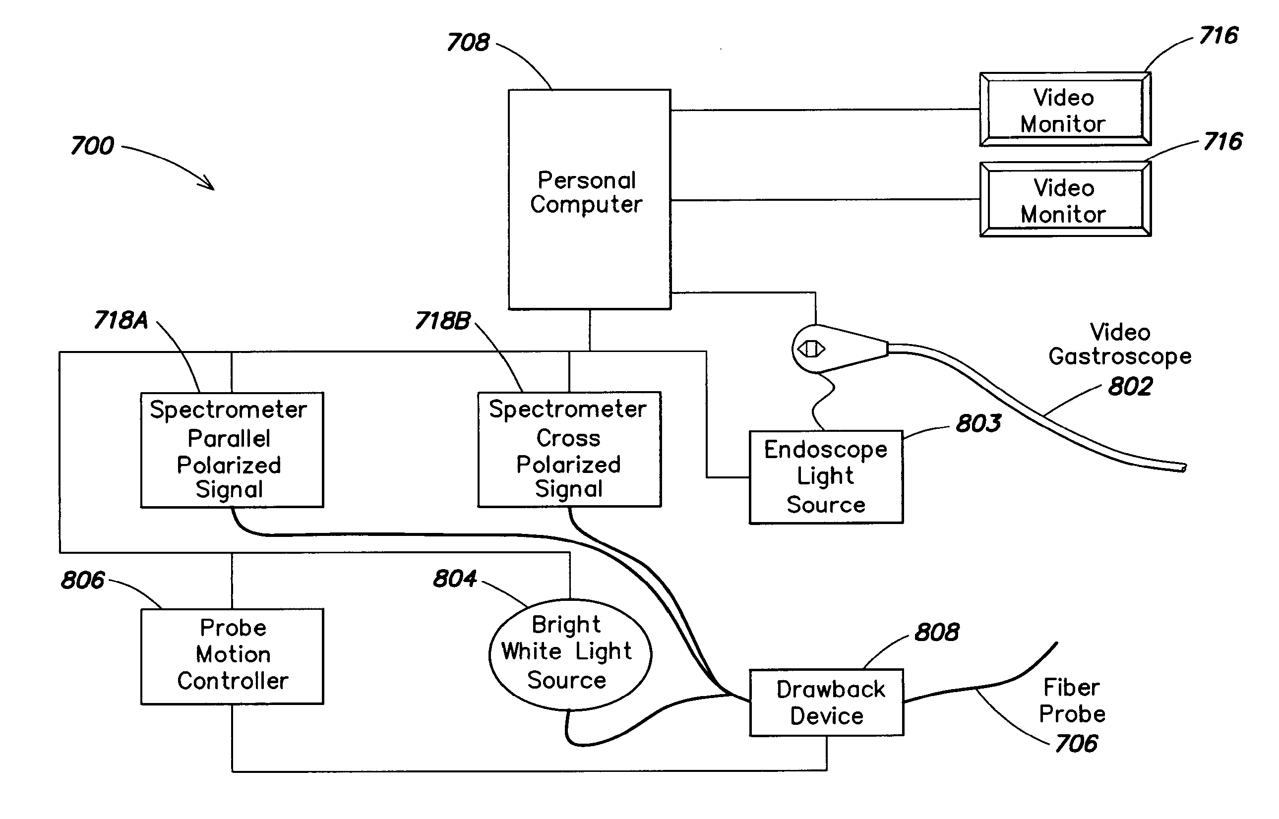
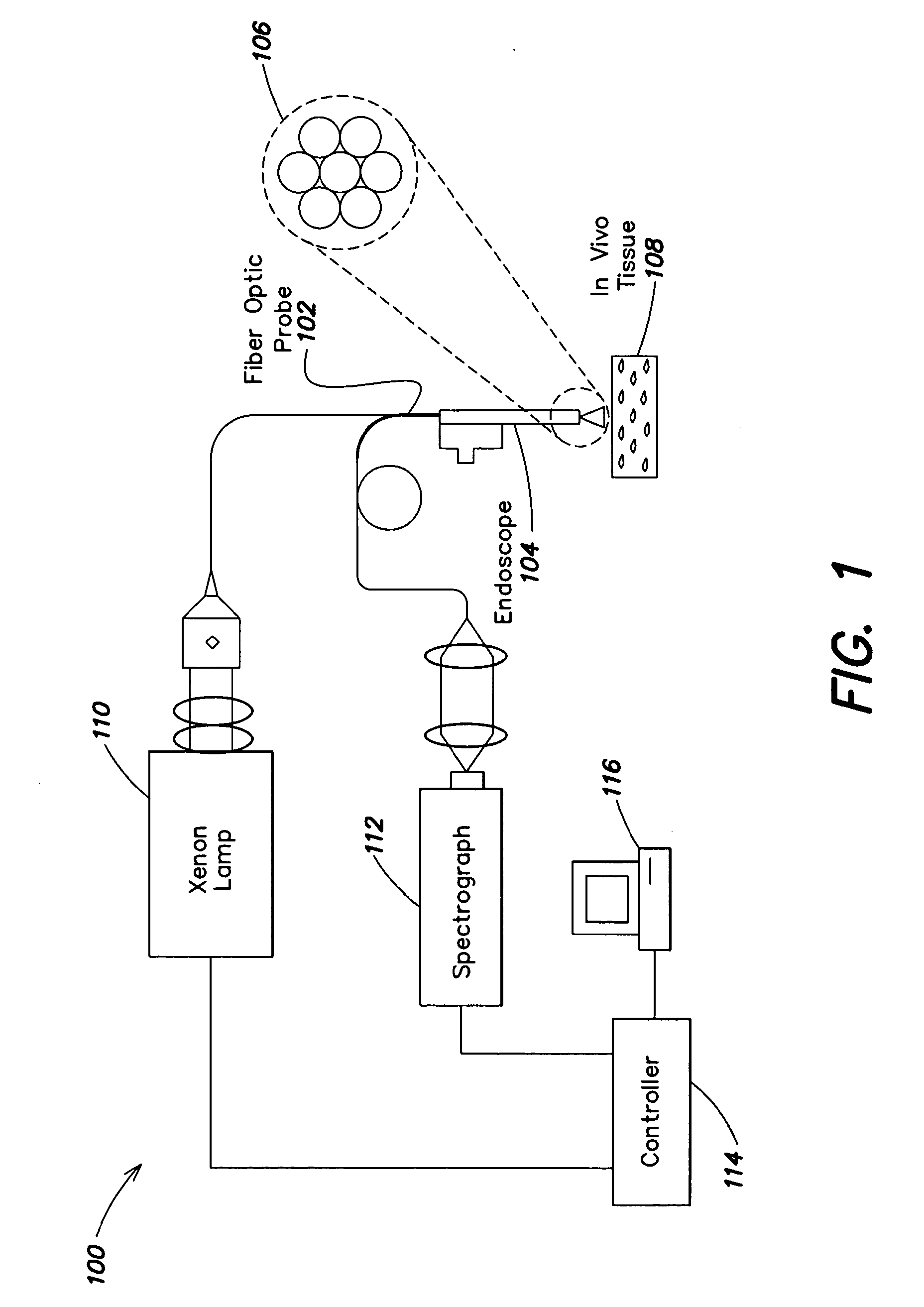
[0046]Embodiments of the invention provide a system and a method of detecting abnormal changes in tissues of organs using a combination of light scattering spectroscopy (LSS) with endoscopy. Employing a probe for performing the light scattering spectroscopy with either existing or specifically designed endoscopic devices results in improved detection of abnormalities in the tissues of organs that may otherwise remain undetected. In particular, this technique may improve early detection of certain morphological and histological changes which, albeit being potentially precancerous, may be amenable to treatments if detected early. Thus, lives of many patients may potentially be saved.

[0047]Some embodiments of the invention provide techniques for early detection of changes in the tissue of the esophagus that may be initial stages of the esophageal adenocarcinoma. Thus, in accordance with one aspect of the present invention, instruments, systems and methods for examining a portion of the...


PUM


Abstract

Method and system for early detection of precancerous and other abnormal changes in tissue of various organs. The system comprises a combination of endoscopic scanning with light scattering spectroscopy and improves detection of abnormalities that may otherwise remain undetected. The system may include a probe that collects data of quality that is independent of a distance of the probe from the scanned tissue. During endoscopy, tissue of an organ is imaged using polarized multispectral light scattering scanning and results are presented to a user in a manner that allows detecting abnormal morphological and biochemical changes in the tissue. A determination of whether to perform biopsy may be performed while the endoscopy is being performed, which thus provides guided biopsy. An entire surface of the organ may be rapidly scanned and results of the scanning are analyzed with a reduced time delay.

Description

RELATED APPLICATIONS[0001]This application claims the benefit under 35 U.S.C. §119(e) of U.S. provisional application 61 / 147,074, filed Jan. 23, 2009, entitled “POLARIZED LSS ENDOSCOPIC SCANNING INSTRUMENT,” the content of which is incorporated herein in its entirety.FIELD OF THE INVENTION[0002]The present invention relates to imaging tissue of an organ using polarized multispectral light scattering scanning method to detect abnormal morphological and biochemical changes indicative of precancerous dysplasia or other tissue abnormalities.BACKGROUND[0003]Early detection of precancerous changes in various organs in the human body may improve results of treatment and ultimately save lives. Techniques used to detect such changes include visual endoscopy and collection of tissue samples (i.e., biopsy) at predetermined sites that are typically used to analyze tissue of organs for abnoinial changes.[0004]As an example, tissue of the esophagus may be examined using light scattering spectrosc...

Claims


Application Information

Patent Timeline
Patent Type & Authority Applications(United States)
IPC IPC(8): A61B5/1455A61B6/00
CPCA61B5/0062A61B5/0084A61B5/0075A61B5/0071A61B5/0066
Inventor PERELMAN, LEV T.
Owner BETH ISRAEL DEACONESS MEDICAL CENT INC
Why Eureka
Try Eureka