Method for 3-dimensional microscopic visualization of thick biological tissues

a biological tissue and microscopic visualization technology, applied in the field of 3d visualization of thick biological tissue, can solve the problems of reducing the field of 3d imaging, and reducing the depth of imaging depth

Inactive Publication Date: 2012-04-05
NATIONAL TSING HUA UNIVERSITY
View PDF4 Cites 9 Cited by
  • Summary
  • Abstract
  • Description
  • Claims
  • Application Information

AI Technical Summary

Benefits of technology

[0006]The present invention discloses a method for 3-dimensional microscopic visualization of thick biological tissues. This method includes: providing a thick biological tissue which is processed by an aqueous optical-clearing solution so as to allow light to travel across the thick biological tissue, providing a removal apparatus (or a cutter) to remove a portion of the thick biological tissue, using an image capturing apparatus to capture the image of a surface of the thick biological tissue, increasing a focal depth of the image capturing apparatus incrementally so as to capture the image stack from surface of the thick biological tissue to the ...

Problems solved by technology

Although the microtome-based 2-dimensional (2D) histology is the current standard method for tissue analysis, diagnostic errors and inconclusive results are embedded in the process due to incomplete sampling—only a few sections of the specimen are examined to represent the bulk tissue.
However, because opt...

Method used


Image

  • Method for 3-dimensional microscopic visualization of thick biological tissues
  • Method for 3-dimensional microscopic visualization of thick biological tissues
  • Method for 3-dimensional microscopic visualization of thick biological tissues

Examples


Embodiment Construction

[0014]The present invention will be described in detail using the following embodiments and it will be recognized that those descriptions and examples of embodiments are used to illustrate but not to limit the claims of the present invention. Hence, other than the embodiments described in the following, the present invention may be applied to the other substantially equivalent embodiments.

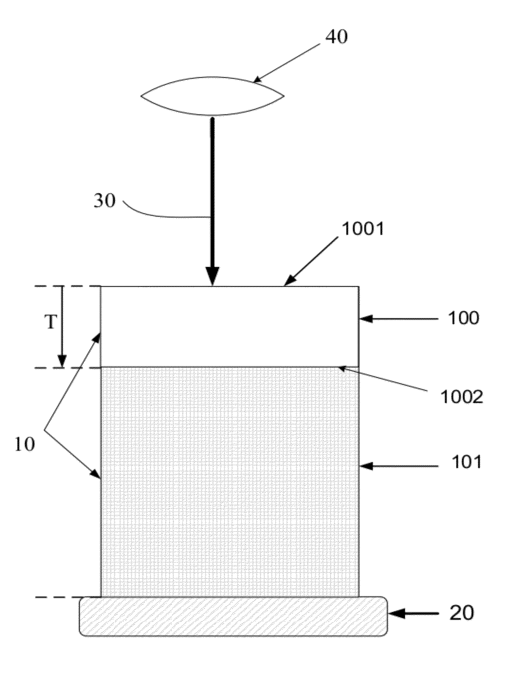
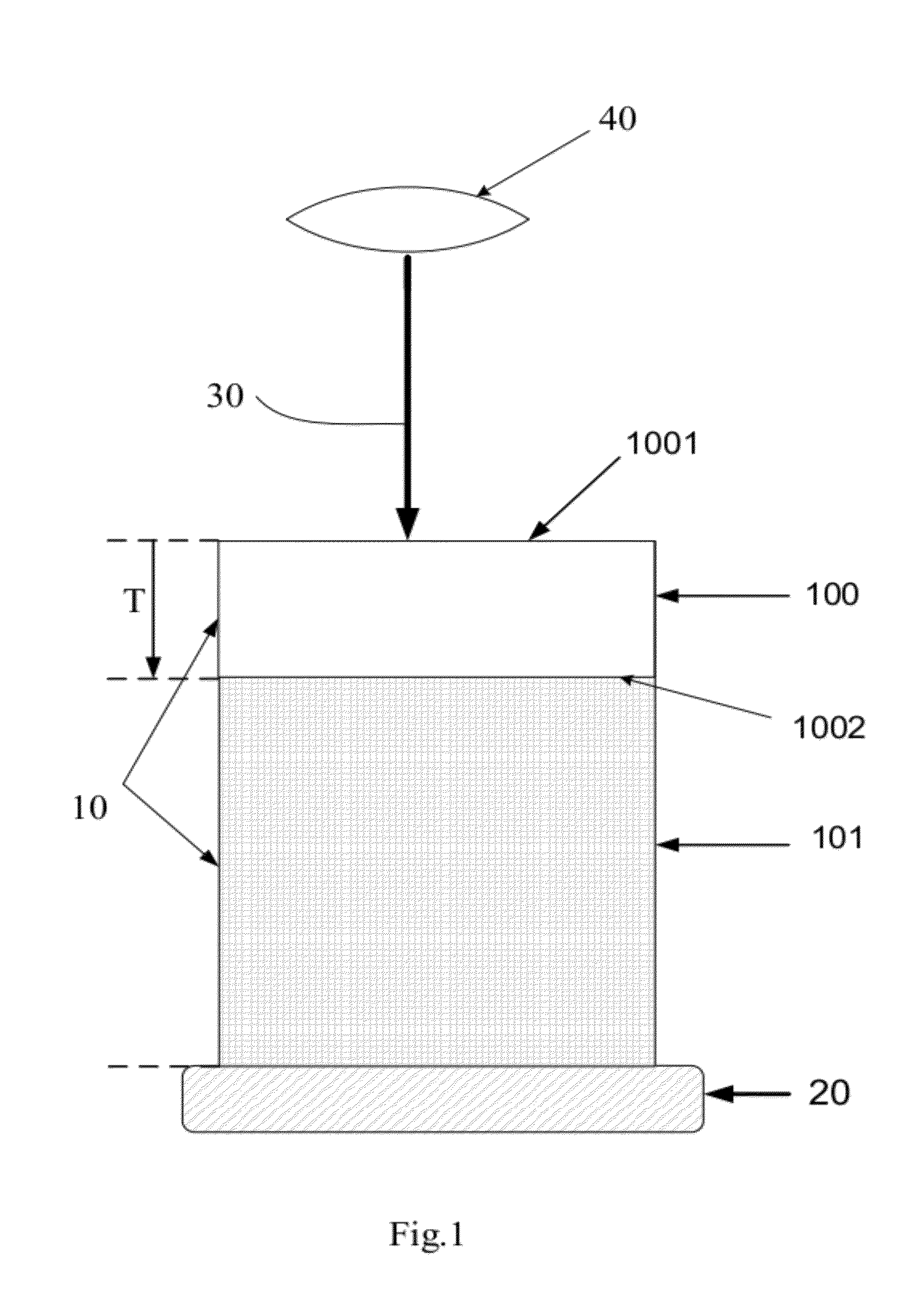
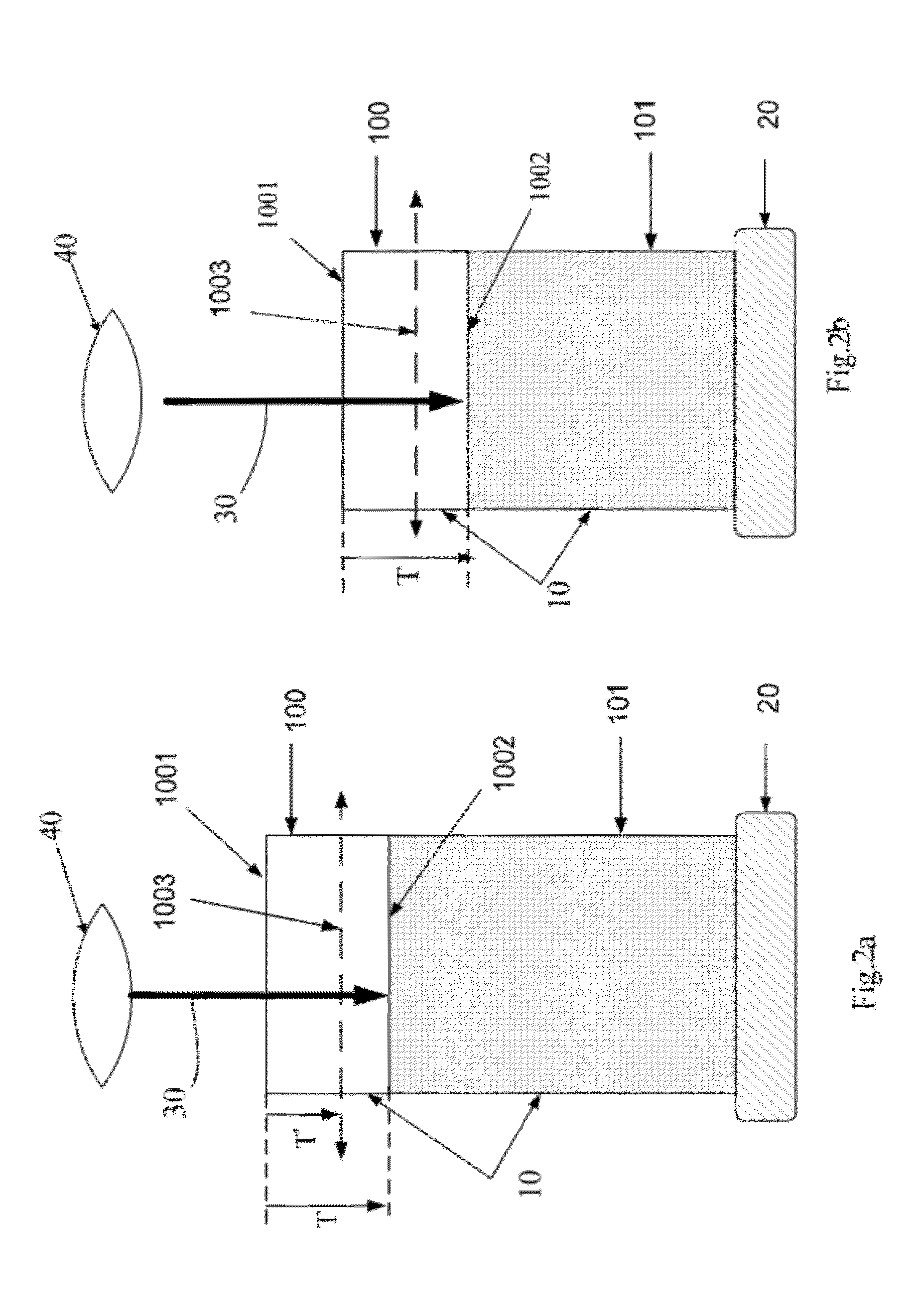
[0015]A method for visualizing the 3-dimensional microstructure of a thick biological tissue is disclosed in present invention. This method includes: utilizing a technique to increase the imaging depth of a tissue, scanning and capturing images of the tissue within the boundary plane where the limitation of imaging depth occurs, defining a removal plane upper than the boundary plane, and removing the tissue above the removal plane. Because the source of light scattering is largely reduced by tissue removal, the second round of imaging acquisition can be performed to extend the imaging depth to new ...


PUM


Abstract

The present invention discloses a method of visualizing the 3-dimensional microstructure of a thick biological tissue. This method includes: a process of immersing thick, opaque biological tissues in the optical-clearing solution, for example FocusClear (U.S. Pat. No. 6,472,216), and utilizing an optical scanning microscope and a cutter. In microscopy, the cutter removes a portion of the tissue after each round of optical scanning. Each round of optical scanning follows the principal that the depth of the removal plane is less than the depth of the boundary plane derived from the scanning This method acquires an image stack to provide the information of thick biological tissue's 3-dimensional microstructure with minimal interference by the tissue removal.

Description

CROSS-REFERENCE TO RELATED APPLICATIONS[0001]This application claims the benefit of TAIWAN Patent Application Ser. No. 099133619, filed on Oct. 1, 2010, which are herein incorporated by reference.TECHNICAL FIELD[0002]This invention relates generally to a method of visualizing a 3-dimensional structure of a thick biological tissue, and more particularly to a method of combining the optical-clearing process (use of immersion solution to reduce random scattering as light travels across media), confocal microscopy, and controlling tissue removal to visualize the 3-dimensional microstructure of a tissue block such as the endoscopic biopsy.BACKGROUND OF THE RELATED ART[0003]Three-dimensional (3D) microscopic visualization of tissue of interest is crucial in biomedical research. For example, 3D visualization of a suspicious area of the patient's biopsy provides a global view of the tissue organization. This feature is particularly valuable when intricate vascular and neural networks are th...

Claims


Application Information

Patent Timeline
IPC IPC(8): H04N13/02
CPCG02B21/006
Inventor LIU, YUAN-ANCHIANG, ANN-SHYNTANG, SHIUE-CHENG
Owner NATIONAL TSING HUA UNIVERSITY
Why Eureka
Try Eureka