Shear flow device and methods of use
a technology of shear flow and flow device, which is applied in the direction of measuring device, material analysis through optical means, instruments, etc., can solve the problems of large sample volume, high cost associated with large high-quality pieces of quartz needed to construct such a cell, and the device used for partially orientating molecular samples is considerably complicated, etc., to achieve convenient maintenance and adaptability, easy to find quartz plates, and sufficient quality
- Summary
- Abstract
- Description
- Claims
- Application Information
AI Technical Summary
Benefits of technology
Problems solved by technology
Method used
Image
Examples
example 1
Design of the Orientating Device
Prototype and Test Experiments
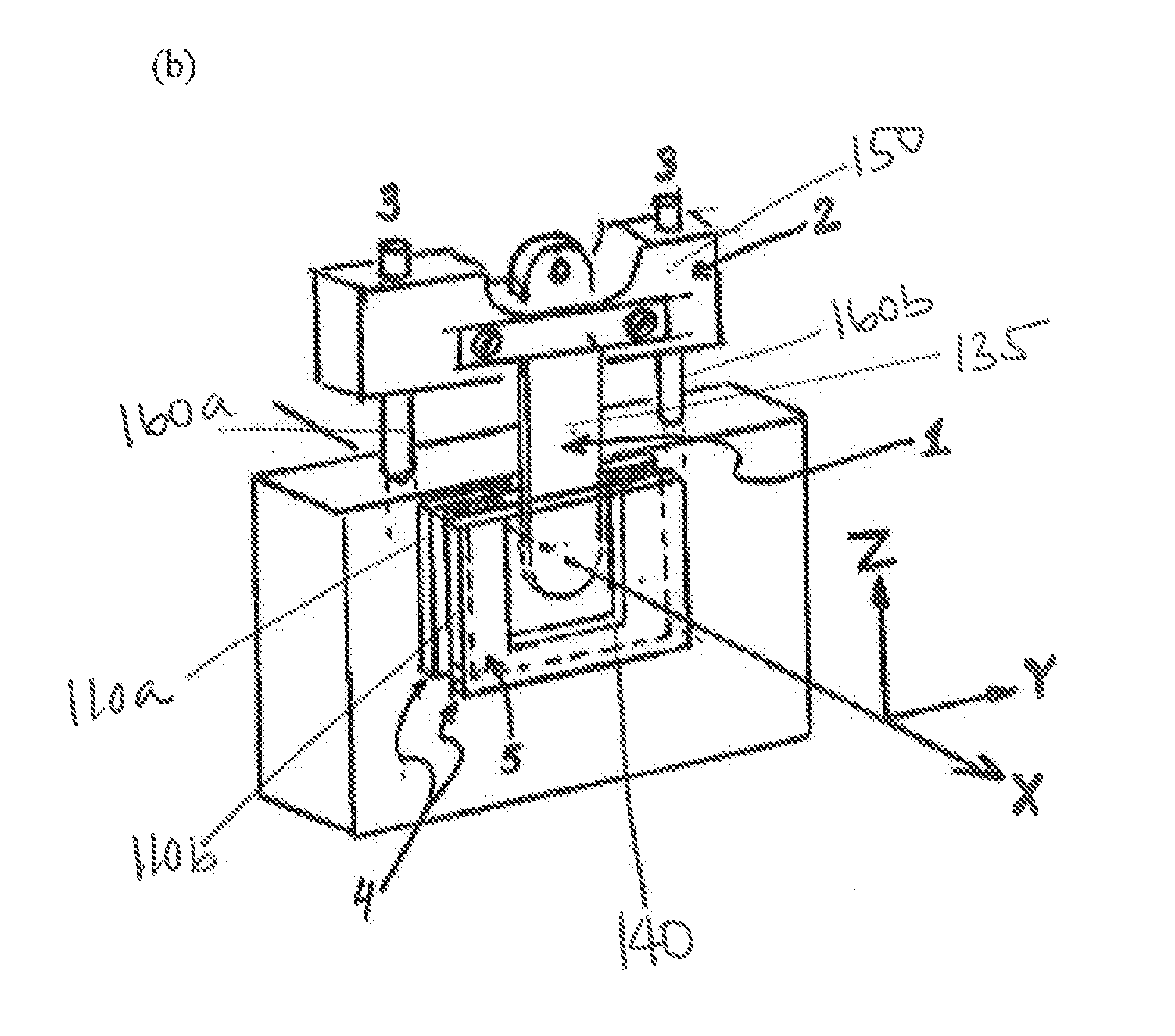
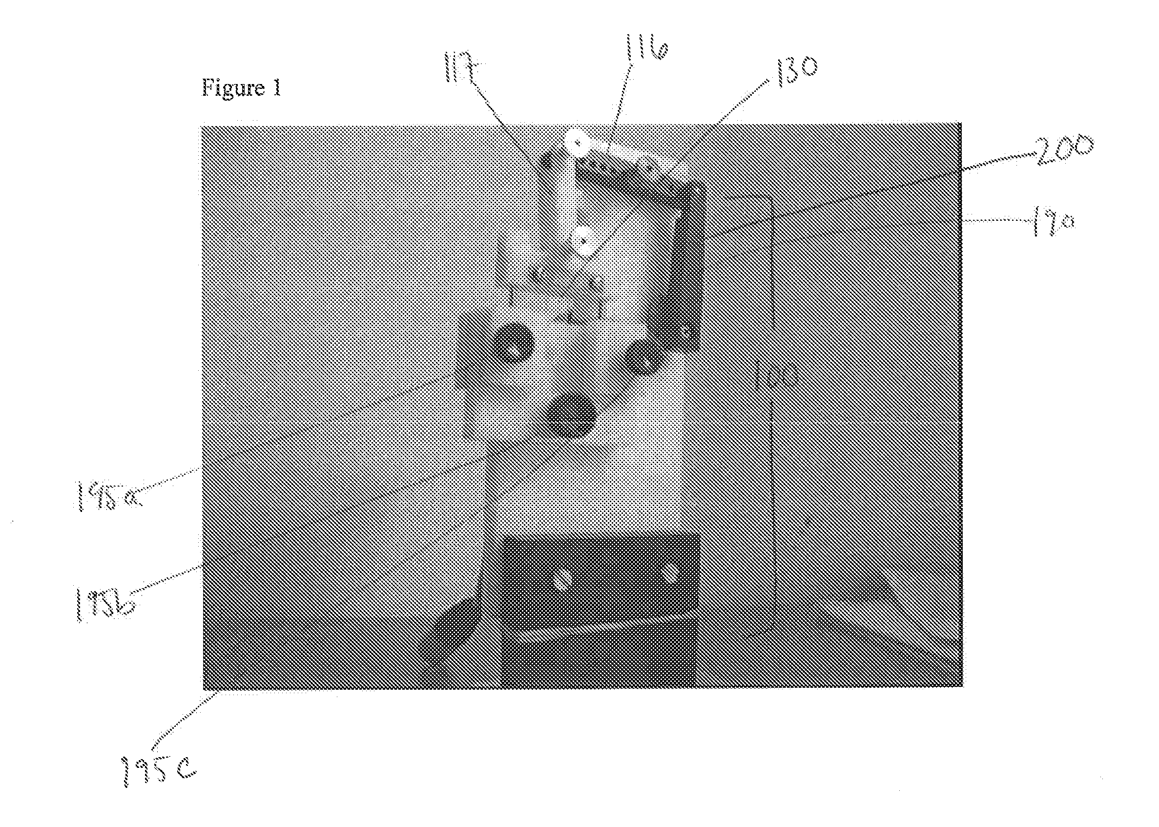
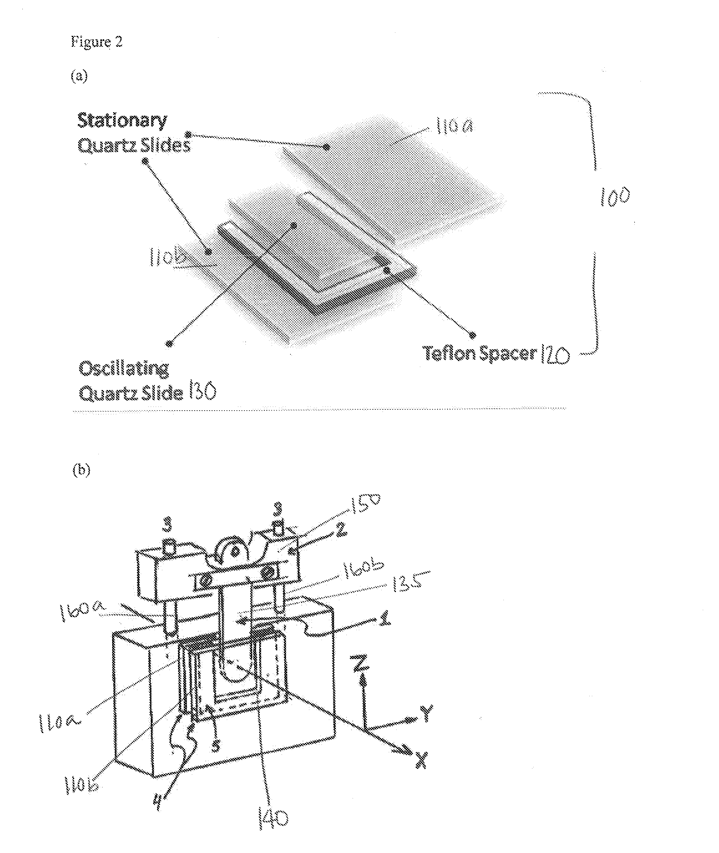
[0090]A machine drawing of the prototype device is shown in FIG. 1. The device is made of fused silica and consists of one static part, the cell, and one moving part, the slide, which has dimensions to fit snugly inside the cell. The slide is connected to a lever that is connected to a micro motor via an eccentric wheel. The motor causes the slide to oscillate in a sinusoidal motion at typically 4-10 Hz and with amplitude adjustable between 1 and 5 mm. The sample, 0.4 ml of a solution of calf thymus DNA, was placed inside the UV cell, the slide introduced so that sample liquid raised above the horizontal light beam of an LD spectrophotometer. An LD spectrum is recorded during oscillation and a baseline recorded at rest. Also, an absorbance spectrum (A) of the isotropic sample is recorded at rest. The quotient spectrum LD / A provides the desired information about DNA orientation and structure (and in case of bound ligand, b...
example 2
Calf-Thymus DNA
[0097]The LD spectra of DNA has been previously examined 1 (18-20). The absorption spectrum is dominated by the Π-Π* transitions of the purine and pyrimidine bases with a peak at 259 nm. In FIG. 3 the raw spectra, baseline and baseline corrected spectra of the ct-DNA is shown. The baseline (dashed) is very straight with only a slight decline towards 185 nm which most likely is due to some absorption of the fused silica. With most Couette devices an uncomfortably intense background LD provides baselines that vary strongly with wavelength, as a consequence of long optical pathlengths through quartz details and with quartz that due to curvature etc may have intrinsic birefringence (due to tensions in the material) which changes the polarization of the light. The LD-absorption spectra is positive since the DNA is oriented vertically in the design. Because the baseline is almost entirely flat, the baseline correction becomes mostly a matter of “zeroing” the spectra.
[0098]F...
example 3
Large Unilamellar Vesicles (LUVs)
[0099]The resulting LD-absorption spectra from pyrene and retinoic acid in LUVs is shown in FIG. 5. In pyrene the two orthogonal transition moments can be seen, with the negative peak at 274 nm as an indication of the Syy orientation, and the positive bands as the orientation in Szz. The negative band in the retinoic acid spectrum at approximately 425 nm on the other hand is thought to be a result of retinoic acid forming dimers and aligning along the surface of the membrane. This would explain the negative sign, and the absorption of such dimers would be expected to be red shifted compared to the single molecule. The LDr for retinoic acid is 0.011 which is approximately 20-25% of what has been achieved in previous studies using a Couette flow cell (6).
PUM
 Abstract
Description
Claims
Application Information
 - R&D
- Intellectual Property
- Life Sciences
- Materials
- Tech Scout
- Unparalleled Data Quality
- Higher Quality Content
- 60% Fewer Hallucinations
Browse by: Latest US Patents, China's latest patents, Technical Efficacy Thesaurus, Application Domain, Technology Topic, Popular Technical Reports.
© 2025 PatSnap. All rights reserved.Legal|Privacy policy|Modern Slavery Act Transparency Statement|Sitemap|About US| Contact US: help@patsnap.com


